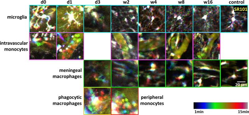
Figure 6. Morphology and dynamics of different types of CX3CR1-GFP cells over time.

Representative temporal-code two-photon images of different types of CX3CR1+ cells at 50μm from the prism surface. Yellow: intravascular dye (SR101). Rainbow spectrum: temporal code of 15 min. Control images were from a non-implant thin skull CX3CR1-GFP mouse. First row (cyan outline): microglia became activated with enlarged cell body and shortened processes in the first several days post implant, and that ramified microglia with thin and motile processes and small soma returned around w2 to w4. Second row (magenta outline): small and round monocytes circulating in the blood vessels after microprism implantations. Third row (green outline): meningeal macrophages were absent at the beginning due to the removal of meninges. They appealed back at 3 DPI and lined up at the brain surface since w2. Forth row: giant foam-like phagocytic cells showed up from ~3 DPI and disappeared after w2 (yellow outline). Small and round monocytes were found wandering in the peripheral corticospinal fluid space around w2 (red outline).
