
FIGURE 5.
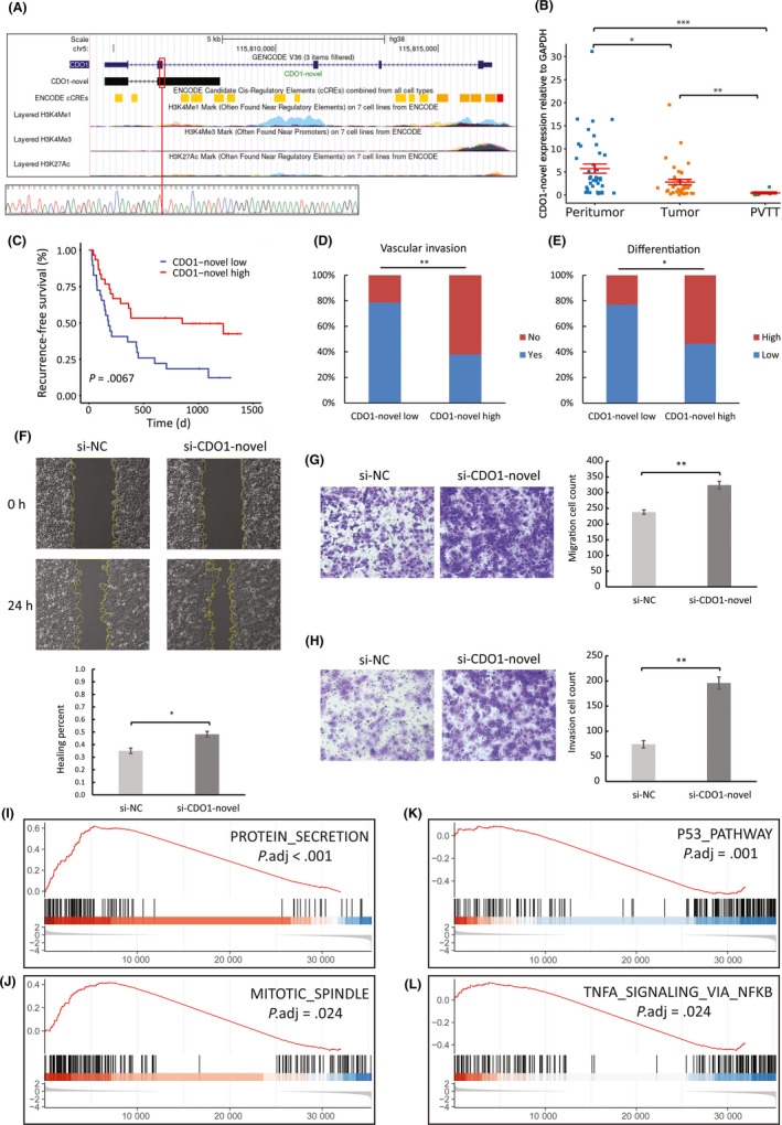
Expression profiling and biological functions of CDO1‐novel in hepatocellular carcinoma (HCC). A, Comparison between annotated and novel CDO1 transcripts, and associated epigenetic data obtained from ENCODE. The novel exon‐retained‐intron junction is denoted by a red vertical line and was validated by Sanger sequencing. B, Quantitative RT‐PCR analysis of CDO1‐novel expression in peritumor (n = 41), tumor (n = 39), and portal vein tumor thrombus (PVTT) (n = 7) of a different cohort of 42 HCC patients (one‐way ANOVA and Tukey’s post‐hoc test). C‐F, Association of CDO1‐novel expression in tumor with prognoses and clinicopathological features using data of the 59 patients. F, Wound healing migration assays of negative control (si‐NC) and CDO1‐novel silenced (si‐CDO1‐novel) cells (n = 3). Transwell migration (G) and invasion (H) assays of si‐NC and si‐CDO1‐novel cells (n = 3). I‐L, Selection of the top enriched hallmark gene sets from Molecular Signatures Database (MSigDB) analyzed with Gene Set Enrichment Analysis (GSEA). The y axis indicates enrichment scores, hits, and ranking metric scores from top to bottom. *P < .05, **P < .01, ***P < .001