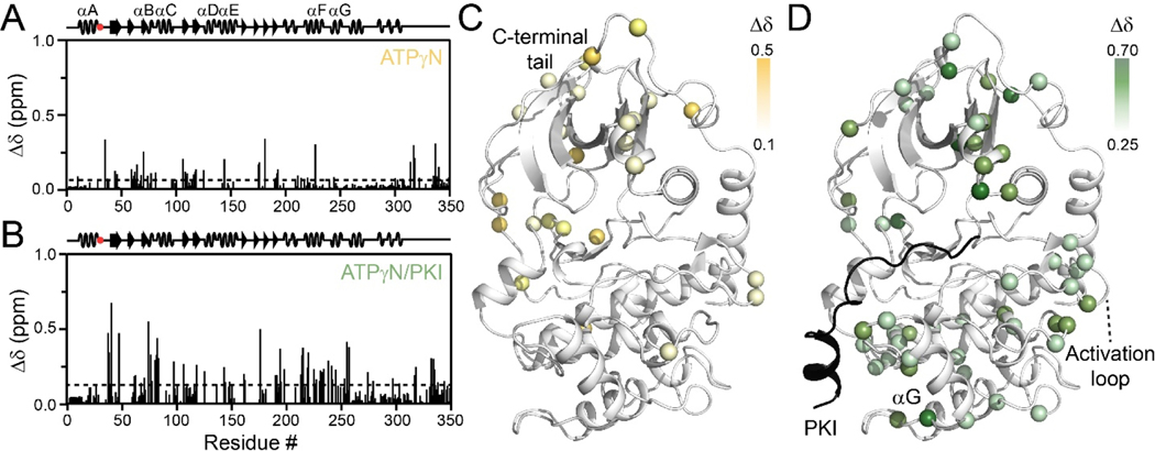
Figure 2. Structural response of PKA-CE31V binding to nucleotide and protein kinase inhibitor.
Chemical shift perturbation (CSP) of amide fingerprint of PKA-CE31V upon binding (A) ATPγN and subsequent binding of (B) PKI. The average CSP is shown as a dashed line. CSPs of PKA-CE31V amide resonances mapped onto the structure (PDB: 1ATP).

