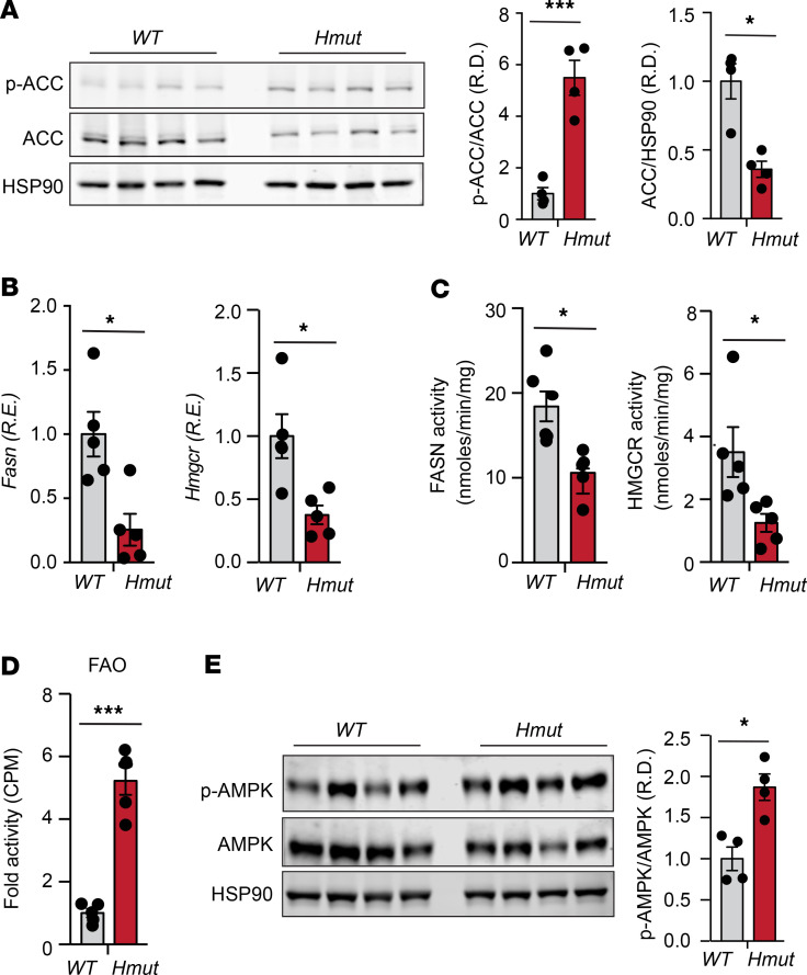
Figure 6. Loss of ANGPTL4 in hepatocytes inhibits DNL pathway and promotes FAO.
(A) Representative immunoblot images showing p-ACC and ACC levels in the liver isolated from fasted WT and Hmut mice fed a CD for 8 weeks. The right panel shows p-ACC/ACC and ACC/HSP90 ratios from immunoblot image quantification. (B–D) Expression and respective activity of the enzymes FASN and HMGCR and FAO in liver isolated from fasted mice. (E) Immunoblot images of p-AMPK and total AMPK protein in liver isolated from fasted mice. Densitometric analysis shown in the right panels. All data are represented as mean ± SEM. *P < 0.05; ***P < 0.001, comparing Hmut with WT mice using unpaired t test.

