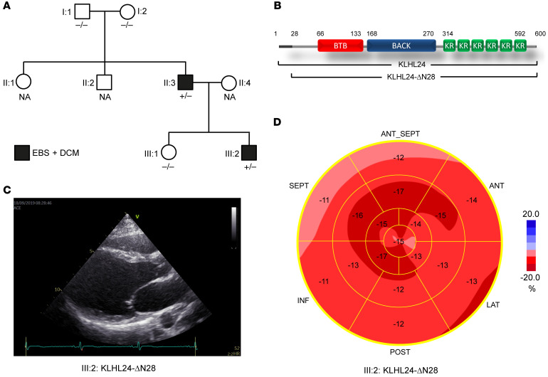
Figure 1. Family tree and clinical characteristics of DCM and EBS.
(A) Family tree showing KLHL24 genotype-phenotype correlations, where +/– indicates KLHL24WT/c.1A>G genotype (heterozygous carrier for mutation c.1A>G in KLHL24) and –/– indicates normal KLHL24WT/WT genotype. NA indicates unknown genotype. Black boxes are members who have an EBS and dilated cardiomyopathy phenotype. (B) Schematic figure depicting the protein domains of KLHL24 and the shorter KLHL24-ΔN28 mutant. BTB: BR-C, ttk, and bab; BACK: BTB and C-terminal Kelch; KR: kelch repeat. (C) Echocardiographic image (long axis parasternal view) of the left ventricle of patient III:2. End-diastolic volume was increased (91 mL/m2; normal value < 75) and ejection fraction was reduced (0.51; normal value > 0.55). (D) Bull’s eye representation of echocardiographic analysis of different regions of the left ventricle with longitudinal strain imaging (normal value –20%) of patient III:2. The apex (central part) is relatively spared; in particular, the basal regions of the left ventricle (outer ring) show decreased strain values, indicating loss of contractile function. The family tree in A was updated after our initial observations (1).

