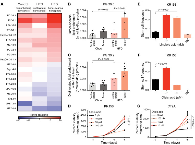
Figure 2. Consumption of HFD drives intracerebral lipid accumulation, promoting tumor cell viability and self-renewal.
(A) Heatmap representing the top 10 most abundant and the bottom 10 least abundant lipids of 216 total lipid species identified by untargeted lipidomic analysis. Four groups (HFD-fed, tumor-bearing hemisphere; HFD-fed, contralateral hemisphere; chow-fed, tumor-bearing hemisphere; and chow-fed, contralateral hemisphere) were compared; n = 5 specimens per group. Heatmap data were normalized to the chow-fed, contralateral hemisphere group. Two modes of lipid enrichment were noted. (B) Four saturated lipid species, including phosphatidylglycerol (PG) 36:0, were identified specifically within the tumors of the HFD-fed animals. P value determined by 1-way ANOVA. (C) Nine mono- or di-unsaturated lipid species, including PG 36:2, were identified within the HFD-fed brain and tumor. P value determined by 1-way ANOVA. In vitro treatment of either (D) KR158 or (G) CT2A with the mono-unsaturated fatty acid oleic acid 18:1 increased tumor cell viability in a dose-dependent manner. P value determined by 2-way ANOVA. (E and F) In vitro limiting dilution analysis conducted with KR158 indicated self-renewal enhancement resulting from exposure to excess oleic or linoleic acid. P values were determined using the Walter and Eliza Hall ELDA portal (60).

