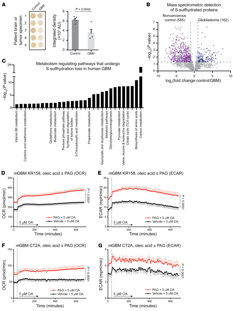
Figure 5. Gliomagenesis induces significant loss in H2S synthesis and signaling primarily associated with cellular metabolism.
(A) Analysis of H2S production confirms that human GBM tumors produce a lower amount of H2S than noncancerous control brain tissues. Each well contains brain or tumor tissue homogenate from separate biopsy specimens. P values determined by unpaired t test. (B) Volcano plot representing the LC-MS S-sulfhydration analysis reveals striking deficits in the posttranslational H2S signaling profile of human GBM as compared with noncancerous human brain tissue. (C) KEGG pathway analysis of the proteins that have undergone S-sulfhydration loss in the context of GBM identifies a broad-spectrum molecular reprogramming centered on GBM tumor cell metabolism. Inhibition of H2S synthesis drives cultured GBM cells into a state of enhanced cellular energetics. Long-term culture of the syngeneic GBM models KR158 and CT2A with PAG resulted in increased metabolic fitness and cellular energetics when compared with vehicle control conditions. Enhanced metabolism was evident at baseline and persisted after introduction of the fatty acid substrate oleic acid. Assessments of cellular metabolism and energetics were based on mitochondrial respiration (D and F), measured by the rate of oxygen consumption (OCR) as well as cellular glycolysis (E and G), measured by the extracellular acidification rate (ECAR). Seahorse Analyzer experiments were conducted in biological triplicate. P values determined by 2-way ANOVA.

