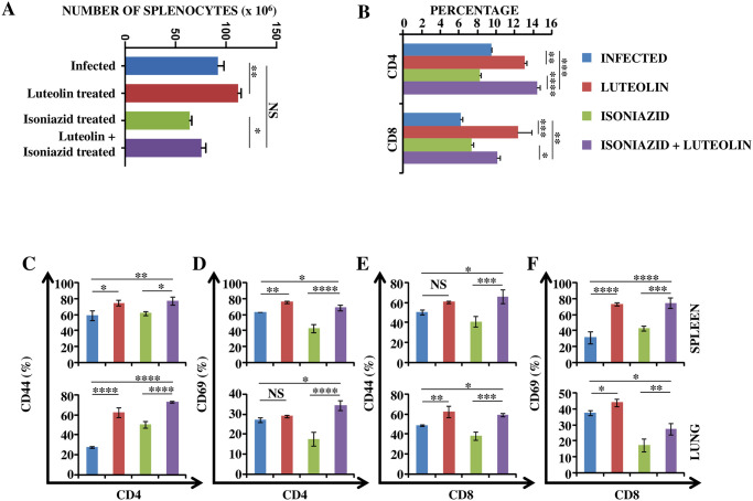
Fig 2. Luteolin induces protective T cell responses.
T lymphocytes isolated from lungs and spleens of the indicated groups of experimental mice at 60 days post-infection were surface-stained with anti-CD4, -CD44 and -CD69 antibodies on ice and fixed prior to acquisition by flow cytometry. (A) Number of splenocytes in Infected, Luteolin-treated, Isoniazid-treated and Luteolin+Isoniazid-treated mice. (B) Percentage of CD4+ and CD8+ T cells in splenocytes of Infected, Luteolin-treated, Isoniazid-treated and Luteolin+ Isoniazid-treated mice. CD4+ T cell activation shown by CD44 (C) and CD69 (D) in spleen (upper panel) and lung (lower panel) of mice infected with H37Rv and treated with Luteolin, INH or Luteolin+INH. CD8+ T cell activation shown by CD44 (E) and CD69 (F) in spleen (upper panel) and lung (lower panel) of mice infected with H37Rv and treated with Luteolin, INH or Luteolin+INH. Data shown are representative of three independent experiments with three mice in each group and represent the MEAN±STDEV values. Differences were considered significant at P<0.05 and are represented by * p<0.05, ** p<0.01, *** p<0.001, **** p<0.0001 whereas non-significant differences are denoted by (NS).

