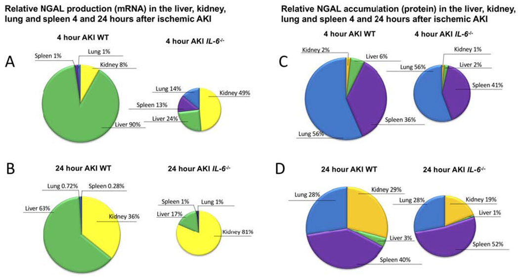
Figure 5. Relative NGAL production (mRNA) and accumulation (protein) after ischemic AKI in wild type (WT) and IL-6−/− mice.

(A,B) NGAL production (mRNA) in liver, kidney, lung, spleen is expressed as % organ production and demonstrates that the liver is the major organ source of NGAL production 4 and 24 hours post-AKI in WT mice after ischemic AKI; in IL-6−/−, the kidney is the major source of NGAL production. Thus, IL-6 is a mediator of hepatic, but not renal, NGAL production. (C, D) NGAL protein levels in the liver, kidney, lung, and spleen are expressed as % organ levels and demonstrate that after AKI, NGAL accumulates predominantly within the kidney, lung, and spleen (total protein was determined within organs, thus, organ accumulation does not distinguish free NGAL from NGAL contained within neutrophils). A similar distribution of NGAL accumulation was observed in IL-6−/− mice. (The size of the pie chart were adjusted to indicate relative production and accumulation between WT and IL-6−/−.) The differences in mRNA and protein highlight that while the liver is the primary site of organ production, NGAL accumulation is widely distributed.
