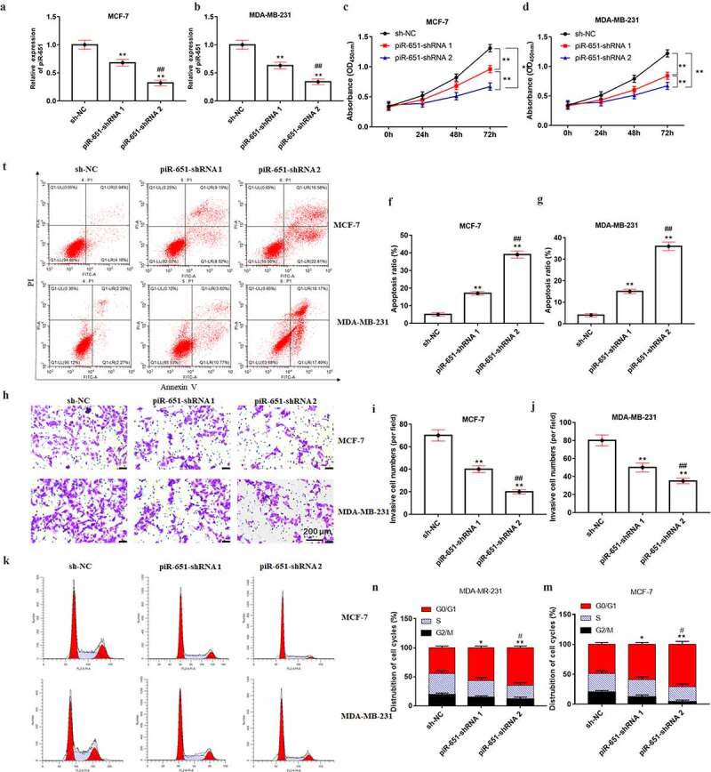
Figure 3.

Interference piR-651 expression inhibits cell proliferation and invasion and promotes apoptosis. Different doses of piR-651 shRNA and its negative control were transfected into MDA-MB-231 and MCF-7 cells for 24 h. (a) RT-qPCR was used to perform the expression of piR-651 in MCF-7 cells. (b) RT-qPCR was used to perform the expression of piR-651 in MDA-MB-231 cells. (c) The cell viability of MCF-7 cells was detected by CCK-8 assay. (d) The cell viability of MDA-MB-231 cells was detected by CCK-8 assay. (e-g) The cell apoptosis ability of MCF-7 cells and MDA-MB-231 was determined by flow cytometry. (h-j) The cell invasion ability of MCF-7 and MDA-MB-231 cells was determined by Transwell assay. (k-m) The percentage of MCF-7 and MDA-MB-231 cells in each phase of the cell cycle was conducted by flow cytometry analysis. N = 4, **P < 0.01 compared with sh-NC group. ##P < 0.01 compared with piR-651 shRNA 1 group. Data were presented as mean ± SEM, (N = 4). The difference of the samples between the two groups were analyzed with independent sample t test. The analysis of variance (ANOVA) test was used to evaluate multigroup comparisons of the means
