Figure 7.
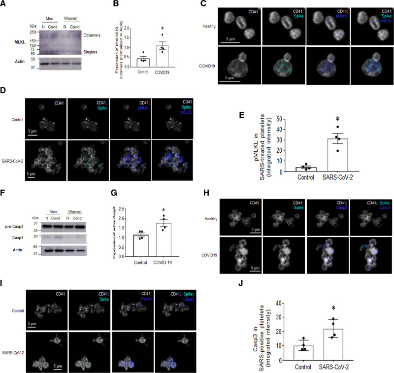
Platelets from patients with coronavirus disease 2019 (COVID-19), and platelets from healthy donors treated with SARS-CoV-2 (severe acute respiratory syndrome, corona virus-2), have upregulated markers of programmed cell death. Washed platelets from patients with COVID-19 were subjected to Western blot (WB) analysis. Markers for necroptosis (A) MLKL (mixed lineage kinase domain-like pseudokinase)-octamer formation by WB (representative images of n=4, 3 males [M], 1 female [F]; the images shown here are from sex- and age-matched groups that include a male and a female pair). B, Quantification of A. C and D, Colocalization of SARS-CoV-2 with p-MLKL (phosphorylated MLKL) by immunofluorescence (IF). C, In COVID-19 blood and in D. Platelets postincubation with SARS-CoV-2 for 30 min. E, Quantitation of D using integrated intensity over platelet number (ImageJ). Marker for apoptosis. F, Active Casp3 (caspase 3) expression (representative images of n=4, 3M, 1F; the images shown here are from sex- and age-matched groups that include a male and a female pair) and its (G) quantitation and H and I. Colocalization of SARS-CoV-2 and Casp 3 in platelets in H. Whole blood from patients with COVID-19 and (I) platelets incubated with SARS-CoV-2 for 30 min by IF; (J) quantitation of I as in E. IF was performed on whole blood that was immediately RBC-lysed and fixed with 4% paraformaldehyde and then stained with the indicated markers (platelet marker used is CD41). IF images are representative of n=4 different patients with COVID-19 on n=4 different healthy donors. Significance was assessed using a nonparametric t test (Mann-Whitney), *P values for B and G are 0.0289; *P values for E and J are 0.0286; data are presented as mean±SEM.

