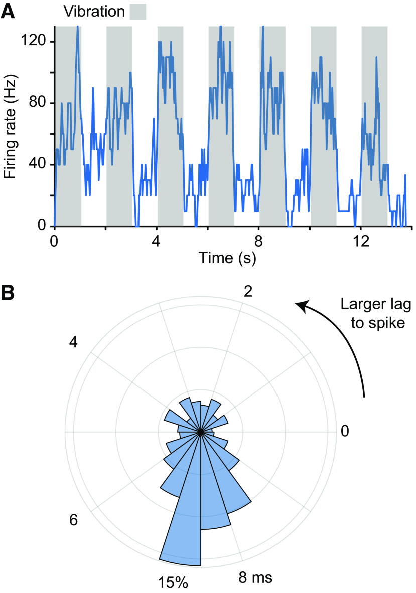
Figure 4.
Responses to muscle vibration of a spindle-receiving neuron. A: response of a CN neuron to 100-Hz vibration applied to the brachialis muscle belly. Gray regions indicate the stimulation epochs. The neuron’s firing rate rose quickly to 100 Hz and returned to baseline immediately when the vibration stopped. B: phase locking between the vibration peaks and action potentials. We computed a phase histogram between the mode voltage applied to the stimulation and evoked spikes. The mode at ∼7.5 ms indicates that the vibration peak led this neuron’s spikes with a reliable latency. Some of the breadth of the peak is certainly due to the sinusoidal nature of the stimulus. CN, cuneate nucleus.

