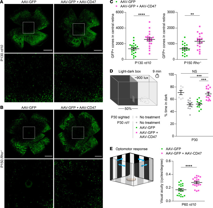
Figure 3. Effect of CD47 expression on long-term cone survival and visual function.
(A) Representative flat mounts of P130 rd10 retinas following infection with AAV8-RedO-GFP or AAV8-RedO-GFP plus AAV8-RedO-CD47. Paired images depict low- and high-magnification views. Scale bars: 1 mm. (B) Representative flat mounts of P150 Rho–/– retinas following infection with AAV8-RedO-GFP or AAV8-RedO-GFP plus AAV8-RedO-CD47. Paired images depict low- and high-magnification views. Scale bars: 1 mm. (C) Quantification of GFP-positive cones in central retinas of rd10 (n = 20) and Rho–/– (n = 18) mice following infection with AAV8-RedO-GFP or AAV8-RedO-GFP plus AAV8-RedO-CD47. (D) Percentage time spent in dark in a 50:50 light-dark box for untreated (n = 7–10) and rd1 (n = 11–13) mice following infection with AAV8-RedO-GFP or AAV8-RedO-GFP plus AAV8-RedO-CD47. (E) Visual thresholds in eyes from P60 rd10 mice (n = 19), as measured by optomotor following infection with AAV8-RedO-GFP or AAV8-RedO-GFP plus AAV8-RedO-CD47. Data are shown as mean ± SEM. **P < 0.01, ***P < 0.001, ****P < 0.0001 by (C and E) 2-tailed Student’s t test and (D) 2-tailed Student’s t test with Bonferroni correction.

