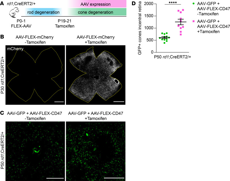
Figure 4. Effect of delayed CD47 expression on cone survival.
(A) Schematic of delayed AAV expression experiments. P0–P1 rd1;CreERT2/+ mice were subretinally injected with FLEX vectors, which were subsequently activated by i.p. injections of tamoxifen from P19–P21. (B) Representative flat mounts of P30 rd1;CreERT2/+ retinas following infection with AAV8-RedO-FLEX-mCherry with or without i.p. injections of tamoxifen. Boundaries of each retina are depicted in yellow. Scale bars: 1 mm. (C) Representative images of central retinas from P50 rd1;CreERT2/+ mice following infection with AAV8-RedO-GFP plus AAV8-RedO-FLEX-CD47 with or without i.p. injections of tamoxifen. Scale bars: 500 μm. (D) Quantification of GFP-positive cones in central retinas of rd1;CreERT2/+ mice (n = 10–12) following infection with AAV8-RedO-GFP plus AAV8-RedO-FLEX-CD47 with or without i.p. injections of tamoxifen. Data are shown as mean ± SEM. ****P < 0.0001 by 2-tailed Student’s t test.

