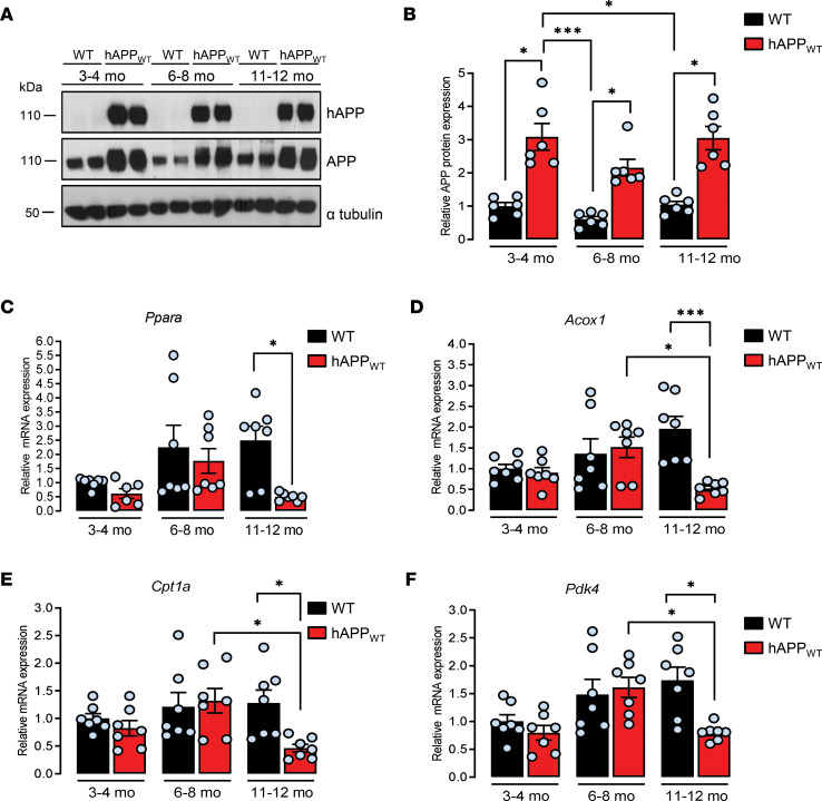
Figure 2. Human APP expression decreases Ppara expression and PPARα downstream target genes in old mice.
Brain frontal cortex tissues from transgenic mice overexpressing nonmutated human APP (hAPPWT) and WT littermates were analyzed at 3–4, 6–8, and 11–12 months old (mo). (A) The expression of hAPP was investigated in mice brain lysates (n = 6 of each) by immunoblot analysis (see complete unedited blots in the supplemental material) with the specific WO2 antibody recognizing hAPP and anti-APP C-terminal antibody recognizing both hAPP and endogenous APP (APP). Blots were further probed using anti–α-tubulin antibody. (B) Relative density of APP expression was compared with α-tubulin. Results were normalized compared with 3–4 mo WT and are shown as mean ± SEM. A Kruskal-Wallis test followed by Dunn’s multiple comparisons posttest was used to assess significance of the mean (APP expression at 3–4, 6–8, and 11–12 mo, P < 0.05). (C–F) Quantitative real-time PCR analyses (n = 7 of each) for Ppara, Acox1, Cpt1a, and Pdk4 mRNA levels. Results were normalized to Rpl32 mRNA, compared with 3–4 mo WT, and shown as mean ± SEM. A Kruskal-Wallis test followed by Dunn’s multiple comparisons posttest was used to assess significance of the mean (11–12 mo hAPPWT mice: Ppara mRNA, P = 0.012, Acox1 mRNA, P = 0.0008, Cpt1a mRNA, P = 0.031; Pdk4 mRNA, P = 0.022), *P < 0.05, ***P < 0.001.

