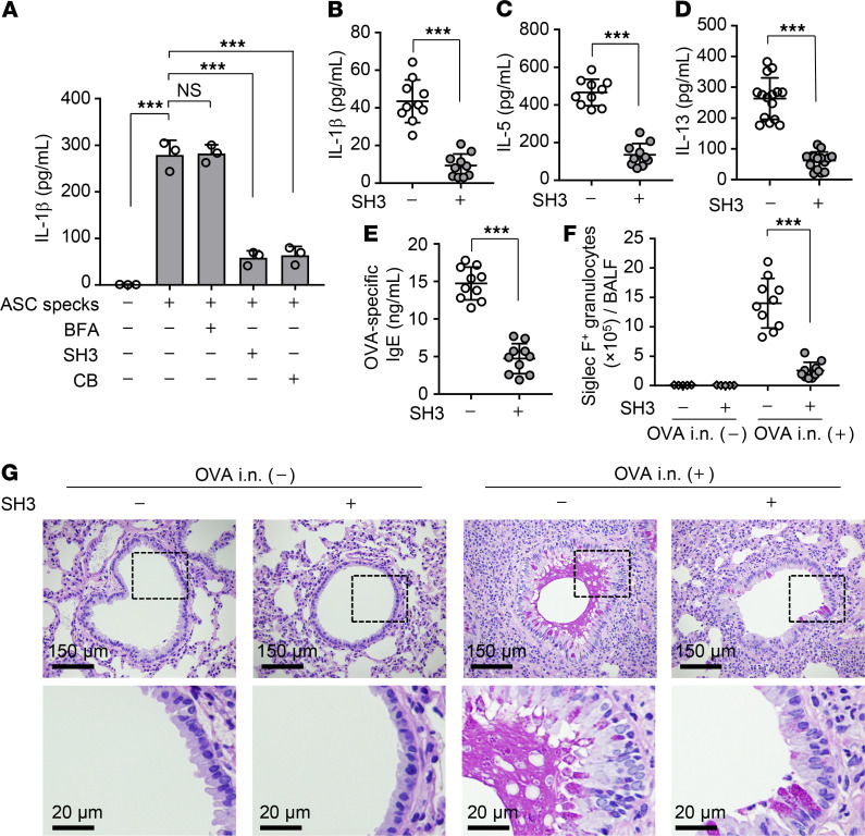
Figure 6. SecinH3 suppresses asthma-like allergic inflammation.
(A) Airway macrophages obtained from WT mice were treated with 100 μM brefeldin A (BFA), 50 μM SecinH3 (SH3), or 30 μM cytochalasin B (CB). At 3 hours after incubation, cells were further incubated with 1 × 10⁴ particles of purified GFP-ASC specks. The level of secreted IL-1β was examined by ELISA at 6 hours after treatment of purified GFP-ASC specks. ***P < 0.001, 1-way ANOVA with Tukey’s test (mean ± SD from 3 independent experiments). (B–G) OVA-immunized WT mice were intranasally injected with OVA at day 7 after the last immunization. After a 1-day incubation, the mice were intranasally administered with 50 nmol/head SecinH3 and then challenged with OVA at days 10 and 13 after the last immunization. The amount of IL-1β (B), IL-5 (C), IL-13 (D), and OVA-specific IgE (E) and the number of Siglec-F–positive granulocytes (F) in BALF were examined at day 1 after the last OVA challenge by ELISA and FACS, respectively (n = 5–10 mice per group). Each symbol represents 1 mouse. ***P < 0.001, 2-tailed Student’s t test. The combined results from 2 independent experiments are shown. Lung tissue sections were stained with PAS-hematoxylin at day 1 after the last OVA challenge (G). Scale bar: 150 μm (top); 20 μm (bottom). Data are representative of 3 independent experiments.

