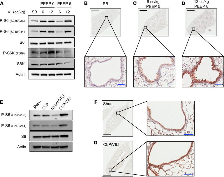
Figure 1. Injurious MV activates mTORC1 in lung epithelial cells.
(A) Immunoblots of phosphorylated and total ribosomal S6, S6 kinase (S6K), and beta-actin using pooled protein lysate from whole lung tissue of SB control mice (n = 3/lane) or mice subjected to MV (n = 4/lane) with high TV (VT, 12 cc/kg), low TV (VT 6 cc/kg), with or without the use of PEEP (0 or 5 cm H2O). Low power (4×) and high power (400×, inset) images from lung tissue that was immunostained for phosphorylated S6 (P-S6, Ser235/236) from SB control mice (B), mice ventilated with noninjurious settings (C) (VT 6 cc/kg, PEEP 5 cm H2O), and mice ventilated with injurious settings (D) (VT 12 cc/kg, PEEP 0 cm H2O). (E) Immunoblots from whole lung tissue of mice subjected to sham laparotomy (n = 3/lane), CLP,(n = 4/lane), and VILI (VT 12 cc/kg, PEEP 2.5 cm H2O) 24 hours after sham laparotomy (n = 3/lane) and VILI 24 hours after CLP (CLP/VILI, n = 4/lane). Representative images from lung tissue that was immunostained for P-S6 (Ser235/236) from sham (F) and CLP/VILI (G) mice. Scale bars: black bars = 2 mm, blue bars = 50 μm.

