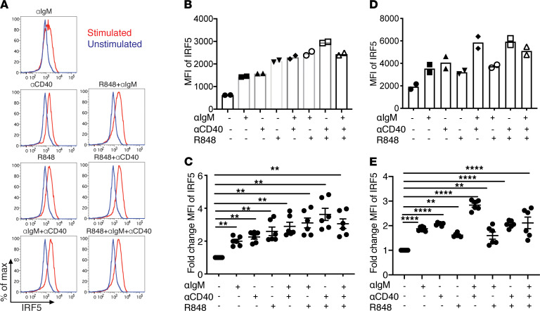
Figure 9. IRF5 expression is increased in activated B cells in vitro.
(A–E) Splenic B cells were isolated from 8- to 10-week-old FcγRIIB−/−Yaa or C57BL/6 mice and were either not stimulated or stimulated with anti-IgM, anti-CD40, and R848 alone or in combination for 24 hours. (A) Intracellular IRF5 levels were measured using flow cytometry. A representative experiment of 6 individual experiments using B cells from FcγRIIB−/−Yaa mice is shown. (B and D) MFI of IRF5 with and without stimulation in B cells from FcγRIIB−/−Yaa mice (B) and C57BL/6 mice (D) (n = 2 per strain). (C and E) Fold change of IRF5 expression normalized to unstimulated control in B cells from FcγRIIB−/−Yaa mice (C) and C57BL/6 mice (E) (n = 6 per strain). Data are shown as mean ± SEM and were analyzed using 1-way ANOVA with Tukey’s post hoc test; **P < 0.01, ****P < 0.0001. IRF5, IFN regulatory factor 5.

