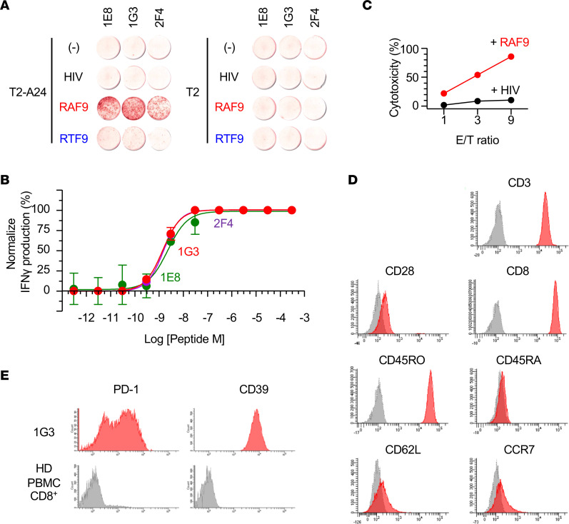
Figure 5. Functions and phenotypes of the RAF9-reactive CD8+ TIL clones.
(A) IFN-γ ELISpot assay of RAF9-reactive CD8+ T cell clones derived from CRC111 TILs (1E8, 1G3, and 2F4) in response to T2-A24 or T2 cells pulsed with 100 nM of the indicated peptides. Data are representative of 4 independent experiments. (B) Functional avidity of 1E8, 1G3, and 2F4 measured by IFN-γ-ELISpot assay against T2-A24 pulsed with a range of indicated concentrations of RAF9 peptide. The number of positive spots was normalized (%). Data represent means ± SEM (n = 3). (C) LDH-release cytotoxicity of 1G3 against T2-A24 cells pulsed with RAF9 or a control peptide at the indicated effector/target (E/T) ratio. The y axis indicates LDH release (%) by target cells. Data are representative of 3 independent experiments. (D) Flow cytometry of 1G3 stained with the indicated antibodies (red) or PBS (gray). (E) Flow cytometry of 1G3 (red) and HD-derived CD8+ PBMCs. Data are representative of 2 independent experiments.

