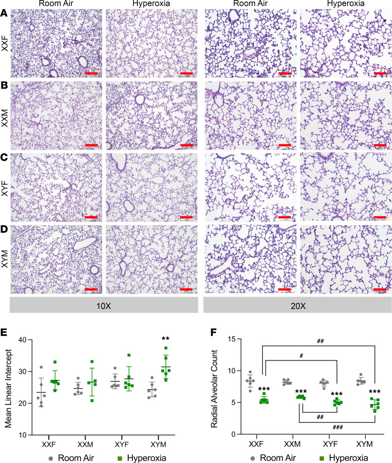
Figure 2. Chromosomal sex but not gonadal sex had a significant impact on alveolarization after neonatal hyperoxia exposure in mice.
(A–D) Representative H&E-stained sections from male and female neonatal mice exposed to room air or hyperoxia (95% FiO2, P1–P4) at ×10 and ×20 magnification in XXF (A), XXM (B), XYF (C), and XYM (D) mice. Lung morphometry in neonatal FCG mice (n = 5–6 mice per group) exposed to hyperoxia (95% FiO2, P1–P4) was assessed using mean linear intercept (MLI) and radial alveolar count (RAC). (E) MLI in FCG neonatal mice exposed to room air or hyperoxia on P21. (F) RAC in FCG neonatal mice exposed to room air or hyperoxia on P21. Data are shown as mean ± SD from 5–6 individual animals. Statistical analysis was performed using 3-way ANOVA to assess the effect of treatment, chromosomal sex and gonadal sex, as well as the interactions between the independent variables. Significant differences between room air and hyperoxia within genotype are indicated by ***P <0.001. Significant differences between hyperoxia-exposed mice between different genotypes are indicated by #P < 0.05, ##P < 0.01, and ###P < 0.001. Scale bars: 200 μm (×10) or 100 μm (×20).

