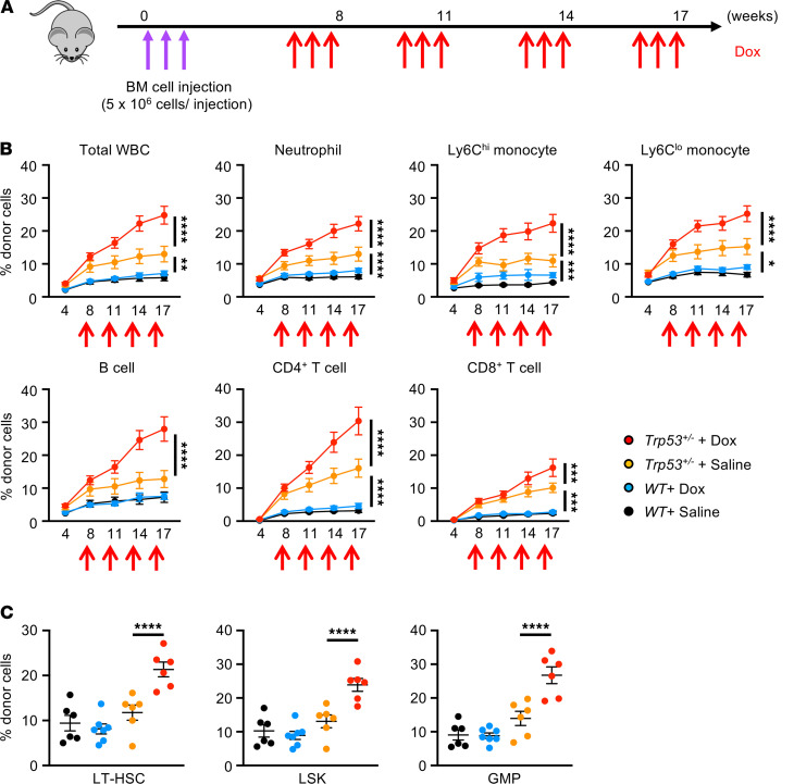
Figure 1. Dox treatment promotes the expansion of Trp53 mutant hematopoietic cells in blood and bone marrow.
(A) Schematic of adoptive bone marrow transplantation and Dox administration. A total of 1.5 × 10⁷ unfractionated donor bone marrow cells were sequentially injected into nonconditioned B6.SJL-CD45.1 recipients over 3 consecutive days (indicated by purple arrows). Donor cells were obtained from either C57BL/6J wild-type mice (Trp53+/+) or Trp53 heterozygous–deficient (Trp53+/–) mice. Recipient mice were injected with Dox at 7 weeks after BMT involving 4 rounds of 6 μg/g i.p. injection (2 μg/g/d over 3 consecutive days), with 3 weeks between each round (indicated by red arrows). Absolute number and the chimerism of test cells in peripheral blood at baseline and after each cycle of Dox or saline administration were evaluated by sequential flow cytometry analysis. (B) Flow cytometry analysis of blood chimerism over the time course to show the progressive expansion of Trp53+/– mutant clones in total WBCs, neutrophils, Ly6Chi monocytes, Ly6Clo monocytes, B cells, CD4+ T cells, and CD8+ T cells after Dox treatment compared with saline administration (n = 6–7 per group). The approximate times of multiple Dox injections are indicated. Statistical analysis was performed with 2-way ANOVA with Tukey’s multiple-comparison tests. (C) Flow cytometry analysis of bone marrow at 19 weeks after adoptive BMT showing increased chimerism (percentage) of donor-derived LT-HSCs, LSKs, and GMPs in mice transplanted with Trp53 heterozygous–deficient cells compared with wild-type cells after 4 rounds of Dox or saline treatment (n = 6–7 per group). Statistical analysis was performed with 2-way ANOVA with Tukey’s multiple comparisons tests. *P < 0.05, **P < 0.01, ***P < 0.001, ****P < 0.0001. LT-HSC, long-term hematopoietic stem cell; LSK, lineage–Sca1+C-kit+ cell; GMP, granulocyte-monocyte progenitor.

