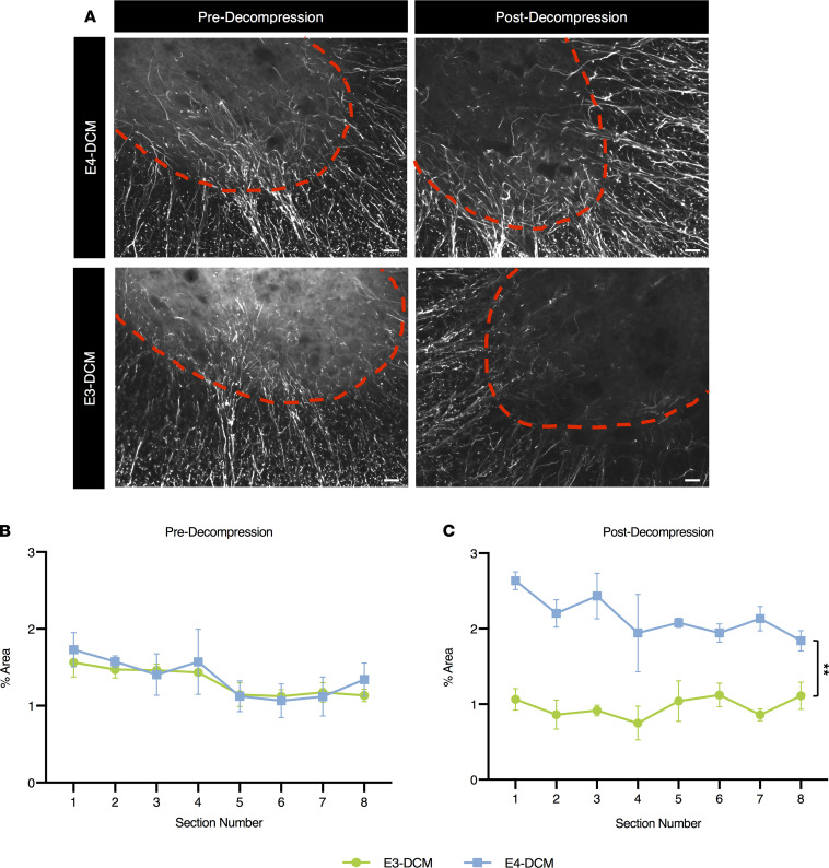
Figure 10. Increased astrogliosis in human ApoE4–knockin mice 24 hours after decompression surgery.
Quantification of GFAP immunoreactivity in the ventral horn region of E3-DCM and E4-DCM mice was normalized to their respective shams. (A) Representative images of the ventral horns for E3-DCM and E4-DCM mice where GFAP immunoreactivity was quantified over an area of constant size. The red dashed line delineates the GM. (B) GFAP immunoreactivity predecompression in E3-DCM and E4-DCM mice showed no significant difference across the 8 cervical spinal cord sections at a 240 μm interval around the injured epicenter. (C) GFAP immunoreactivity in E3-DCM and E4-DCM mice was significantly different postdecompression across the 8 cervical spinal cord sections at a 240 μm interval around the injured epicenter; **P < 0.01. All data are represented as mean ± SEM and analyzed via mixed effects model with Sidak’s post hoc correction for repeated measures. These images are not the same images used for quantification; see Supplemental Figure 2 for representative images. Scale bars: 25 μm.

