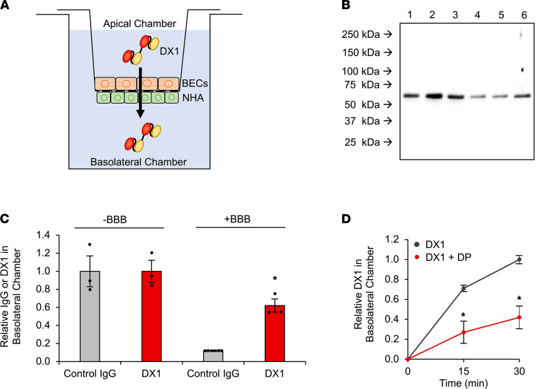
Figure 2. DX1 crosses a transwell model of the BBB in a DP-sensitive manner.
(A) Illustrated transwell model used to test BBB crossing by antibodies. hCMEC/D3 BECs and normal human astrocytes (NHA) were adhered to apical and basolateral surfaces of transwell inserts, respectively. The ability of control IgG or DX1 to cross this model was tested by measuring their appearance in the basolateral chamber after addition to the apical chamber. (B and C) DX1 crosses the transwell model of the BBB. The efficiency of IgG and DX1 transport across control blank inserts and inserts with BBB was compared by anti-IgG or anti-DX1 Western blot of basolateral chamber contents 1 hour after addition of IgG or DX1 to apical chambers. Representative Western blot showing DX1 at expected molecular weight (~54 kDa) in basolateral chambers of blank inserts (lanes 1–3) and BBB inserts (lanes 4–6) is shown in B. Each lane represents an independent experiment. Control IgG blot is shown in Supplemental Figure 2. Presence of the BBB reduced the relative control IgG content in basolateral chambers to 0.12 ± 0.01 relative to chambers lacking the BBB, while DX1 content was only reduced to 0.62 ± 0.07 (P < 0.01 compared with control IgG, Student’s t test, n ≥ 3) as determined by ImageJ-based quantification of band intensities (C). (D) DP inhibits transport of DX1 across the hCMEC/D3 BBB. DX1 content in basolateral chambers was evaluated 15 and 30 minutes after addition of 5 μM DX1 ± 50 μM DP to apical chambers and quantified relative to DX1 content at the 30-minute time point in the absence of DP. *P < 0.01, Student’s t test, n = 3. These data demonstrate DP-sensitive DX1 transport across the hCMEC/D3 transwell BBB model, consistent with ENT2-dependent crossing of the BBB by DX1.

