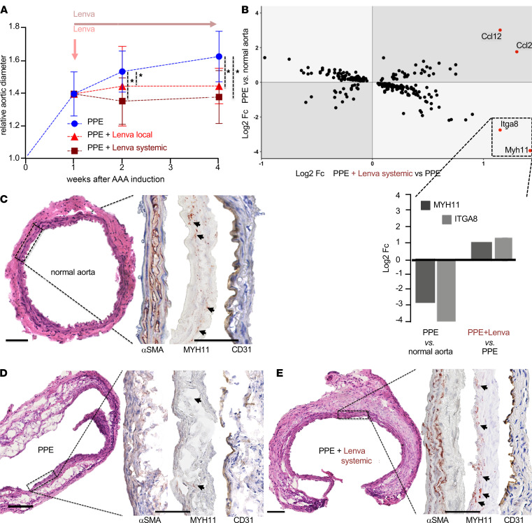
Figure 1. Effects of lenvatinib treatment in vivo.
(A) Two-way repeated measures ANOVA revealed significant effects of time, treatment, and the interaction of time and treatment, on aneurysm diameter in the PPE model. Both systemic oral and local endovascular lenvatinib treatment, starting 7 days after PPE-AAA induction, completely blocked aortic dilatation (local: red, n = 5; systemic: dark red, n = 7) versus sham-treated PPE-AAA mice (blue, n = 11) assessed by B-mode ultrasound imaging (*P < 0.05; displayed relative growth compared with baseline aortic diameter; corresponding absolute diameters shown in Supplemental Figure 1A; P value calculations and detailed measurements in Supplemental Table 1; PPE AAA induction at day 0). (B) Double fold change (Fc) plot depicts analysis of RNA profiling (mouse transcriptome array; MTA_1.0). Areas with light-gray background represent genes regulated in opposite directions (rescue effect) upon lenvatinib treatment. Myh11 displayed the most pronounced changes comparing log2 Fc of gene expression in PPE-induced AAA (PPE) versus control and PPE-induced AAA + lenvatinib (PPE+Lenva systemic) versus PPE. The bar chart highlights and indicates direction (up or down) of the most prominent changes in gene expression from the Fc plot (MTA analysis: n = 6 PPE; n = 7 PPE+Lenva systemic; n = 5 untreated so-called normal aorta). (C–E) H&E staining reveals a disrupted media with pronounced cellular infiltrates in PPE-AAA (D) compared with control aorta (C). (E) Upon systemic lenvatinib treatment, cellular infiltrates were reduced, with a more compact media, a thickened adventitia, and marked fibrosis. In all 3 groups αSMA staining was positive, emphasizing a more linear organized media in control aortas and upon lenvatinib treatment. MYH11 was absent in PPE and highly positive in the media of lenvatinib-treated mice (dark red stain, highlighted with arrows). CD31+ cells (brown stain) indicate intact endothelial lining in all 3 groups, but only in PPE few positive cells are present in the media and adventitia (original magnification 10×/40×; scale bar: 100 μm). αSMA, α–smooth muscle actin.

