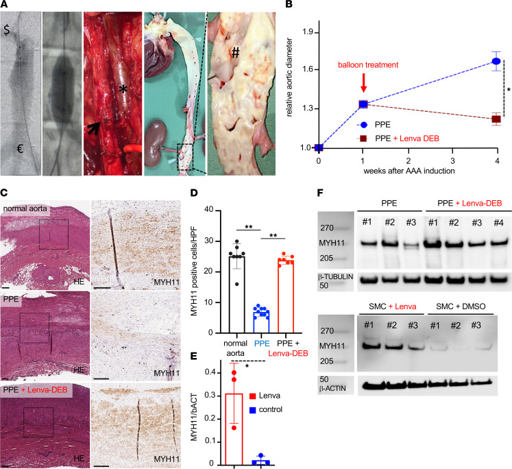
Figure 5. Endovascular lenvatinib treatment LDLR–/– minipigs with AAA disease.
(A) Transfemoral angiography of the infrarenal aorta ($ = right renal artery to € = aortic trifurcation) on day 7 after AAA induction shows the dilated aortic segment. Radiopaque markers guided the DEB (12 × 20 mm) to the lesion. Surgical clips from the aneurysm induction procedure sealed the lumbar vessels. The photograph shows the dilated part of the aorta (arrow) at the time of sacrifice (no circulating blood) next to the cava vein (asterisk). En face preparation of the complete aorta demonstrates severe atherosclerosis and fatty streaks at the level of the aortic valve, the coronary and supra-aortic branch ostia, and the infrarenal level and the renal artery (#). (B) Singular treatment with a lenvatinib-coated balloon (red: Lenva-DEB; n = 4) on day 7 after PPE-AAA induction shows a significant aortic diameter reduction compared with nontreated (blue: PPE only; n = 7) animals (*P < 0.05; 2-way repeated measures ANOVA). (C) H&E indicates a parallel fibrous structure in the nonintervened (normal) LDLR–/– pig aorta with marked noncalcified plaque formation. Similar to the murine PPE-AAA model, a substantial MYH11 restoration was detected upon local lenvatinib treatment (Lenva-DEB) compared with PPE-AAAs. (D) Quantification of MYH11-positive cells confirmed loss of MYH11 in PPE-AAA minipigs compared with untreated (normal) LDLR–/– aorta and restoration upon Lenva-DEB treatment. (E) WB analysis confirmed higher MYH11 protein content in Lenva-DEB–treated minipigs compared with DEB-controls (PPE) with progressing AAA. (F) MYH11 WB analysis on primary pig aortic cells treated for 48 hours with lenvatinib (normalized to β-actin). (*P < 0.05, **P < 0.01; original magnification 5×/20×; scale bar: 200 μm; vessel lumen oriented upward; complete WB and quantifications are shown in Supplemental Figure 13.)

