
Figure 1.
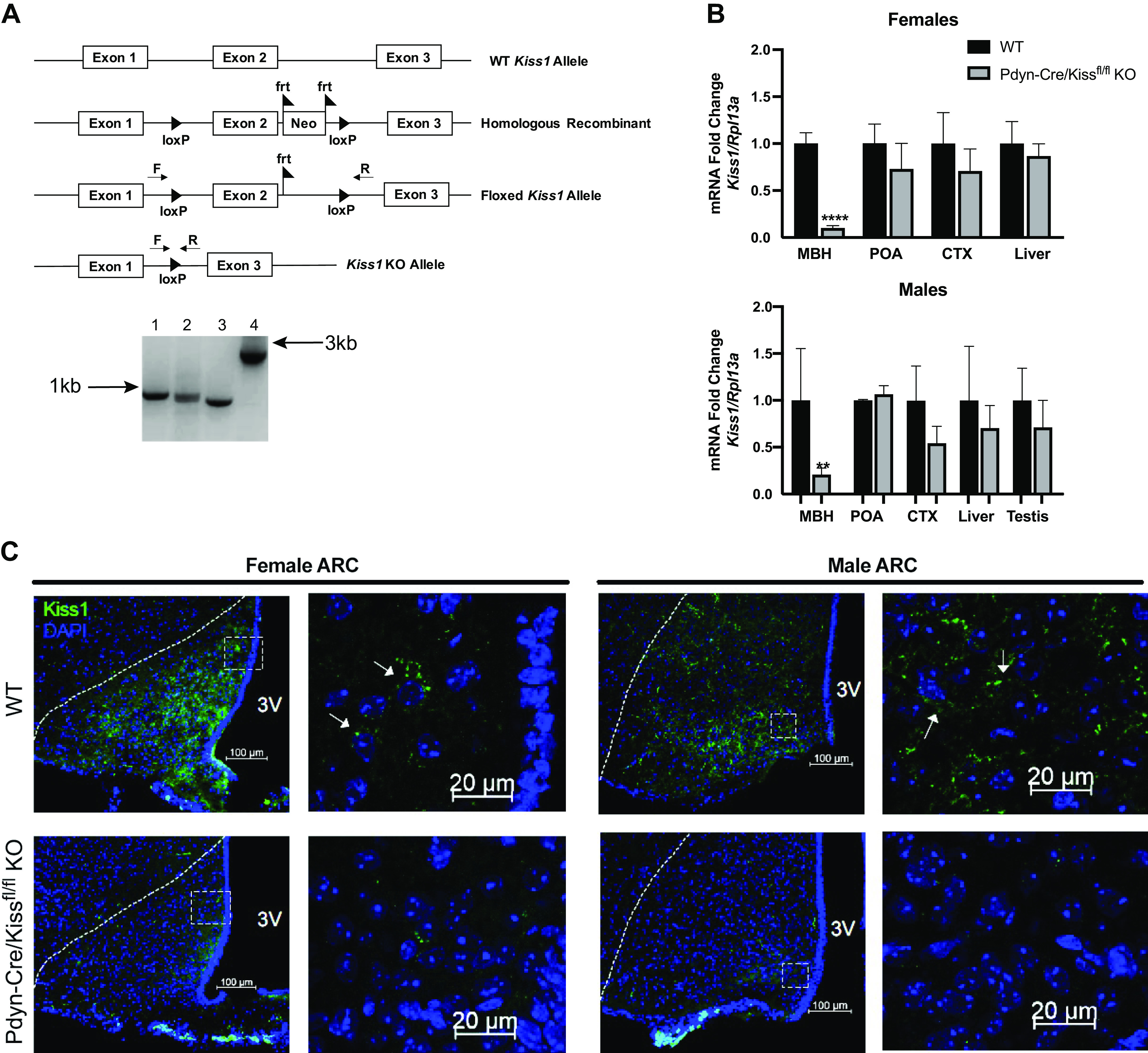
Validation of Pdyn-Cre/Kiss1fl/fl knockout (KO) mouse. A: diagrams of Kiss1 genetic constructs used to generate the Kiss1flox mouse line in the 5′ to 3′ direction. The homologous recombinant expresses the two loxP sites around exon 2 of Kiss1 and the neomycin cassette flanked with frt sites. Postmating a Kiss1 homologous recombinant with a ROSA-FLP mouse, the neomycin cassette was excised, exhibited by the floxed Kiss1 allele. Crossing homozygous floxed Kiss1 mice with Pdyn-IRES-Cre mice resulted in Kiss1 KO alleles in KNDy neurons. Genomic tail DNA was run through a PCR with forward and reverse Kiss1 flox primers that bind outside of the loxP sites. The four allele combinations shown are as follows: 1) flox/flox, 2) flox/wild-type (WT), 3) wild-type/wild-type, and 4) the homologous recombinant including the neomycin cassette and frt sites prior to excision. B: relative mRNA expression of Kiss1 was determined in the mediobasal hypothalamus (MBH), preoptic area (POA), neocortex (CTX), liver, and testes in Pdyn-Cre/Kiss1fl/fl KO animals compared with WT controls within each tissue, within each sex. Kiss1 mRNA in the MBH was significantly decreased in KO mice of both sexes (unpaired Student’s t test, ****P < 0.0001, **P < 0.01 KO vs. WT, (unpaired Student’s t test, WT n = 3–6, Pdyn-Cre/Kiss1fl/fl KO n = 3–6). Kiss1 mRNA in the POA was similar in KO mice of both sexes. 1–3 animals were removed as outliers after conducting Grubb’s outlier test. C: representative photomicrographs revealing kisspeptin peptide immunofluorescence in brain sections from the female arcuate nucleus (ARC) (left, four images) and male ARC (right, four images) of WT and Pdyn-Cre/Kiss1fl/fl KO mice. In the ARC, a decrease in kisspeptin immunoreactivity in neurons in the Pdyn-Cre/Kiss1fl/fl KO mice is seen. Magnification in the leftmost top and bottom panels is set at ×20, while magnification in the rightmost top and bottom panels is set at ×63. Scale bars are set at 100 µm for ×20 images, and 20 µm for ×63 images. Dotted lines in the ×20 image outline the boundary of the ARC. Boxes in the ×20 images denote the frame within which the ×63 images were taken. Arrows denote individual kisspeptin-ir positive cells. F, Kiss1 flox forward primer; Neo, neomycin cassette; R, Kiss1 flox reverse primer, 3V, third ventricle.