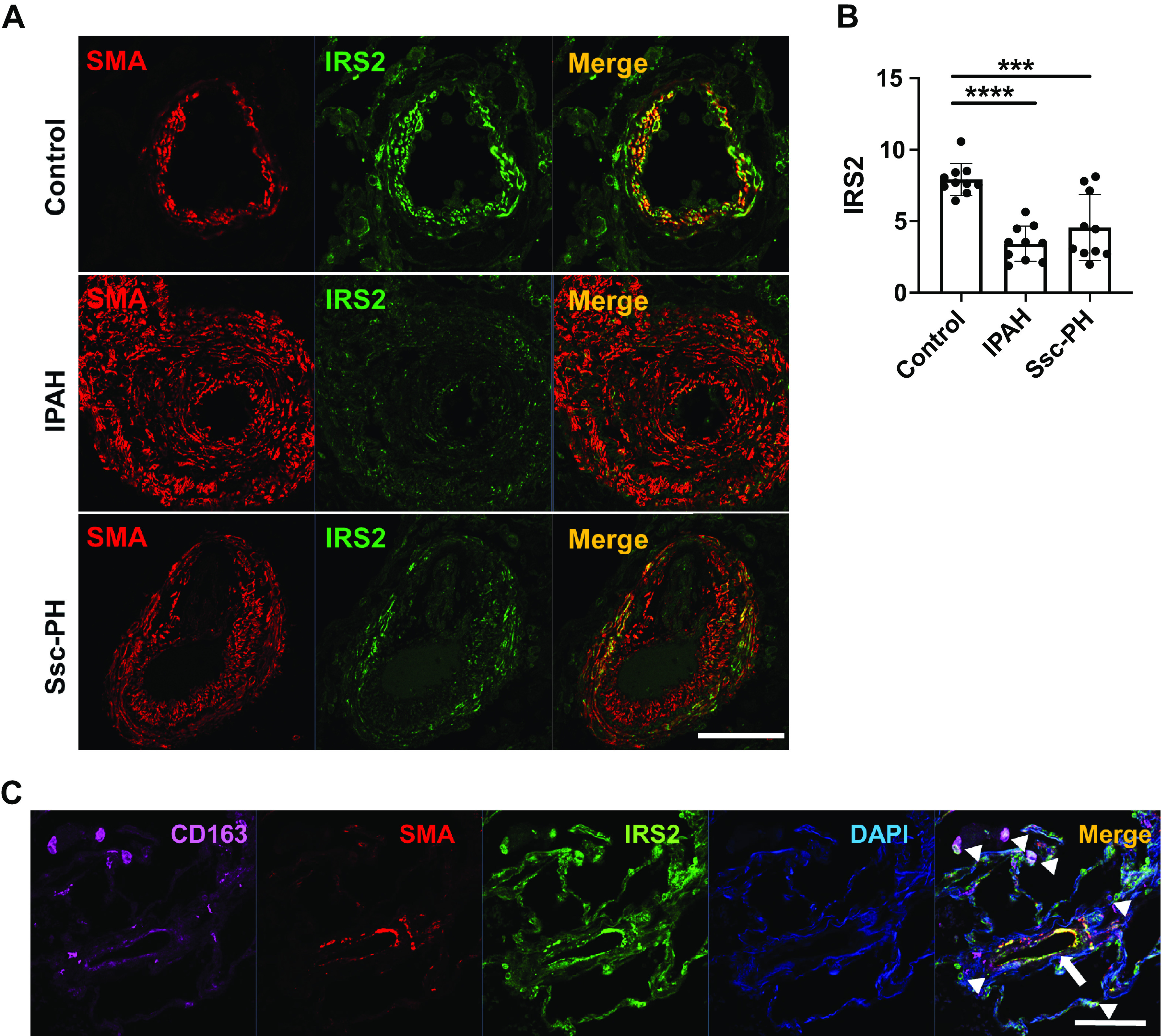
Figure 1.

IRS2 expression is decreased in the pulmonary vasculature (PV) of patients with pulmonary arterial hypertension (PAH). A: expression of IRS2 and smooth muscle actin (SMA) in pulmonary arteries of lung tissue from a normal control subject and patients with idiopathic PAH (IPAH) and scleroderma-associated PAH (Ssc-PAH). IRS2 expression is seen in the PV media and surrounding cells of control lung but is substantially reduced in PAH lung. B: quantification of IRS2 expression in the pulmonary vessels. Fluorescence intensity data are expressed as means ± SD. ***P < 0.001, ****P < 0.0001 (n = 10 vessels/group). Immunofluorescence intensity was significantly diminished in pulmonary vessels of patients with IPAH and Ssc-PAH. C: IRS2-expressing cells colocalize with CD163-positive cells (arrowheads) and SMA-positive cells (arrow). Representative data are shown from normal control subjects (n = 5), subjects with IPAH (n = 5), and subjects with Ssc-PH (n = 5). Scale bars = 100 µm. IRS2, insulin receptor substrate 2.
