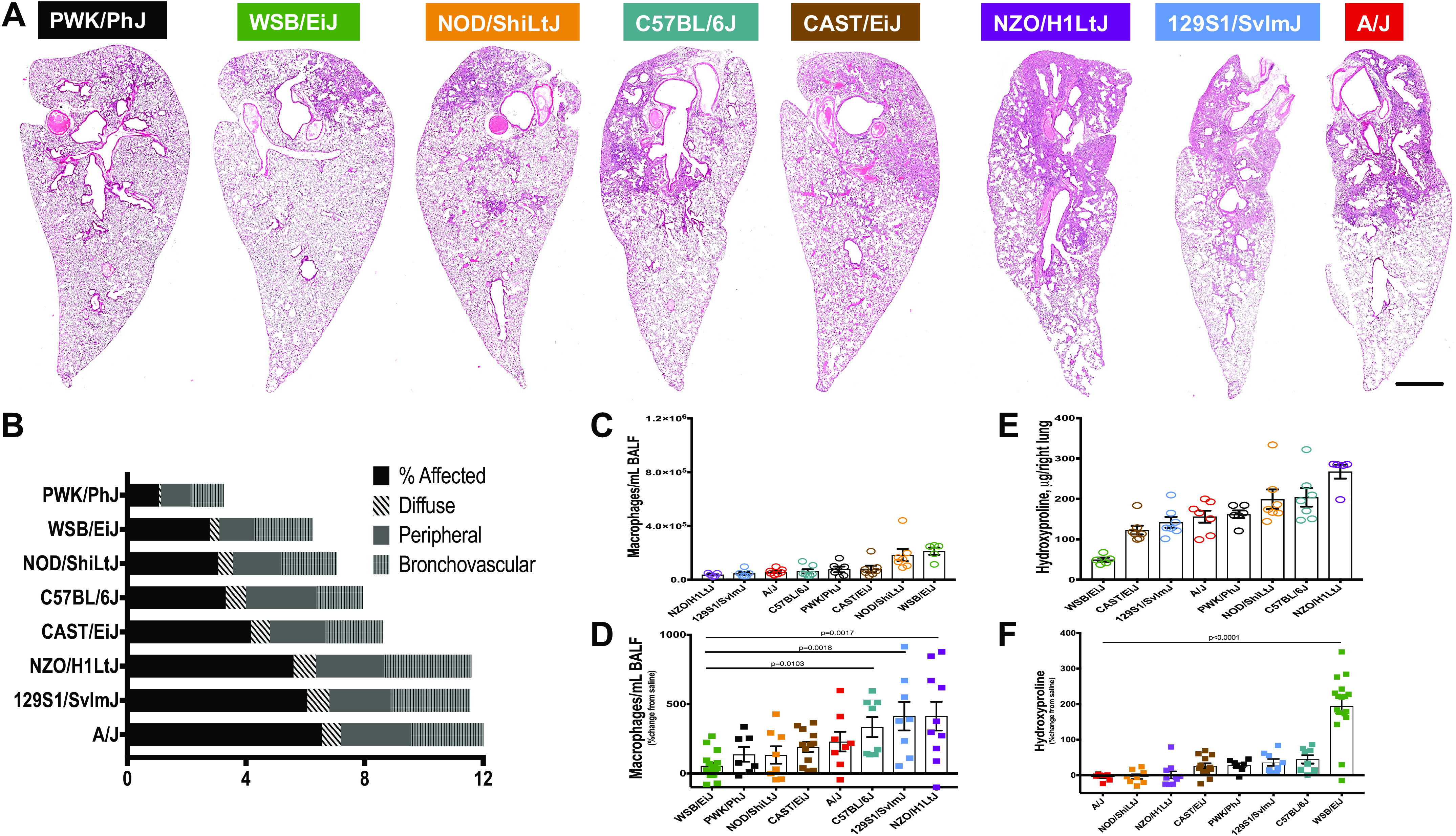
Figure 2.

Comparisons of phenotypes after bleomycin-induced lung injury. Three weeks after a single challenge with intratracheal bleomycin (2.5 U/kg), mice showed considerable variation at baseline and injury. A: representative images of hematoxylin and eosin (H&E) stain across the eight strains of mice show differences in severity and localization of injury. The progressive amount of injured tissue (condensed, darker, with infiltrating immune cells) from PWK/PhJ (almost no injury) to A/J (about 50% of the injured tissue) is shown. Bar is 1 mm. Injury scoring (B); macrophages counts in bronchoalveolar lavage fluid (BALF) (C and D) and hydroxyproline (HP) measurements for total lung collagen (E and F). n = 8–13 mice were analyzed after bleomycin-induced injury and compared with n = 5–7 saline treated-control mice from each strain.
