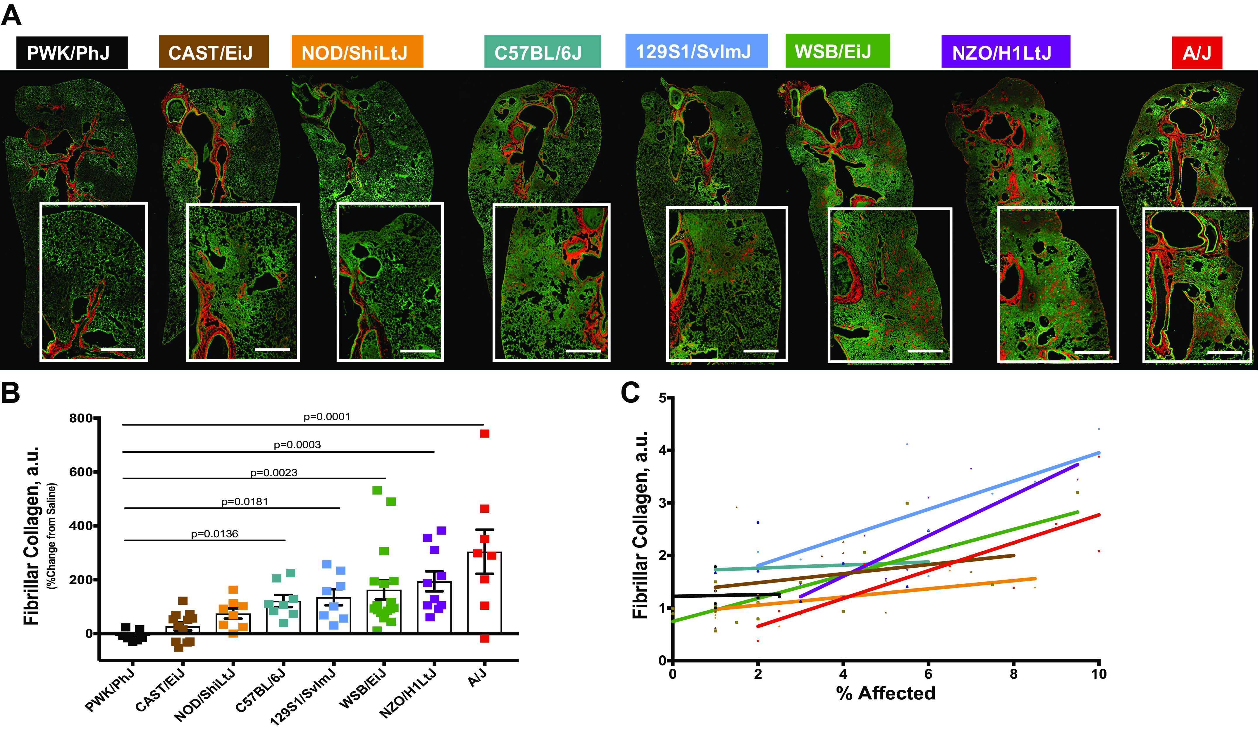
Figure 3.

Second-harmonic generation (SHG) imaging of fibrillar collagen is a more reliable measure of parenchymal fibrosis. Fibrillar collagen was assessed in peripheral lung tissues by confocal/multi-photon fluorescence microscopy with SHG. A: representative images showing the localization of fibrillar collagen (red) around the airways and in the parenchyma following bleomycin exposure. Bar, 500 µm. Fibrillar collagen amount (B) was calculated per mouse following exposure to bleomycin or vehicle. C: fibrillar collagen accumulation correlates with the percent affected area in injured lung. n = 8–13 mice were analyzed after bleomycin-induced injury and compared with n = 5–7 saline treated-control mice from each strain.
