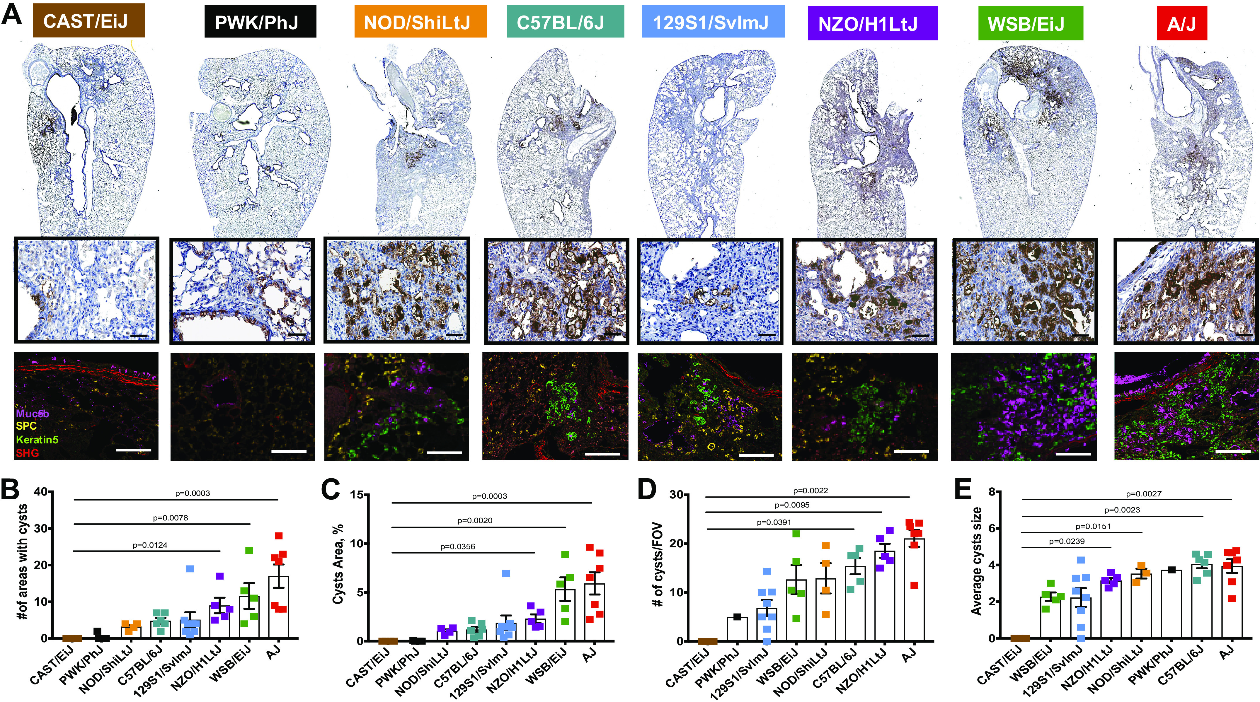
Figure 6.

Muc5b expressing honeycomb-like cysts. A: representative images for Muc5b immunohistochemistry (IHC) staining in eight mouse strains (lower magnification; top row) and regions with cyst-like structures shown at higher magnification (middle row). Immunofluorescent images for representative cyst-like regions with Muc5b (purple), proSPC (yellow), keratin5 (green), and SGH (red) signals in all analyzed mouse strains (bottom row). Bar, 60 µm. Quantification for the number of cyst-like structures (B), area % taken by cyst-like structures (C), number of cyst-like structures in fields of view (FOV; D), and average cyst-like structures size (E). n = 8–13 mice were analyzed after bleomycin-induced injury and compared with n = 5–7 saline treated-control mice from each strain.
