
Figure 4.
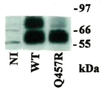
Western blot analysis of WT and Q457R mutant SGLT1 expressed in oocytes. Seven days after injection with WT or Q457R mutant cRNA, protein was extracted from 2 oocytes each. The equivalent to 1/3 oocyte was run in a 12% SDS-PAGE gel and after transfer to a nitrocellulose membrane, and was probed with an SGLT1 antipeptide antibody raised to residues 602–613, at 1:1000 dilution. Both WT and Q457R mutant proteins ran as 2 broad bands, one 70 kDa band which corresponds to the complex glycosylated form, less intense in the mutant, and another of ∼60 kDa which corresponds to core glycosylated form.11,23