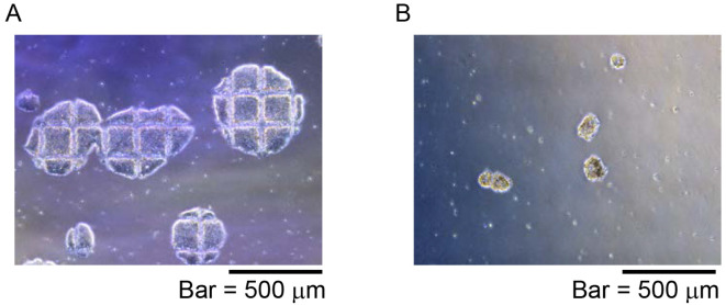
Figure 4. Representative NKT-iPSC morphology for iPS-NKT differentiation.
NKT-iPSC were divided into a few clumps using EZ-passage tool and pipetting. A. Colony shape of NKT-iPSC after cut using StemPro EZ-passage tool; B. Small NKT-iPSC clumps were prepared and collected by pipetting and passage into OP9 feeder cells.

