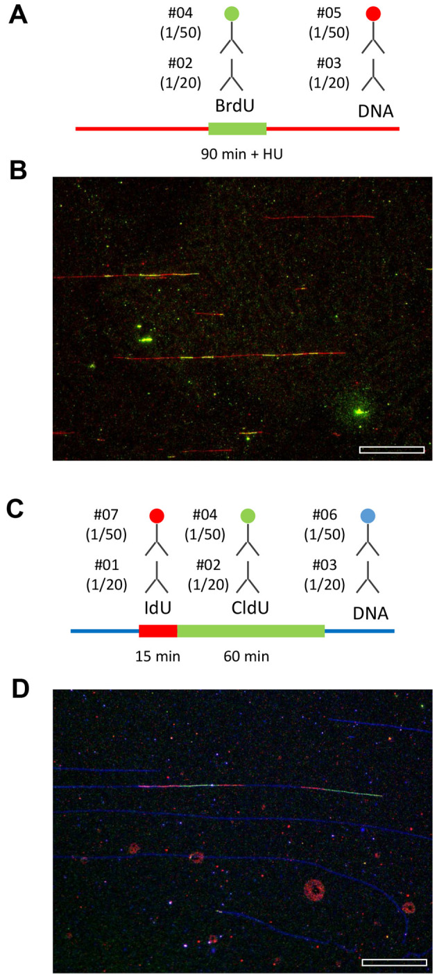
Figure 4. Representative examples of replication patterns observed by DNA combing.

A and B. Yeast cells were released from G1 in the presence of 200 mM HU and labeled with BrdU for 90 min. DNA fibers were purified and stretched by DNA combing. BrdU and ssDNA were detected as indicated. A representative field of view is shown. Bar is 50 kb. C and D. Human cells were labeled for 15 min with IdU and 60 min with CldU. The combination of antibodies used to detect IdU, CldU and ssDNA is indicated. A representative field of view is shown. Bar is 50 kb. Image acquisition is performed with a motorized Leica DM6000B microscope equipped with a CoolSNAP HQ CCD camera (6.45 µm/pixel) and a 40x oil immersion objective. With this setup and a DNA stretching of 2 kb/µm, one pixel corresponds to (6.45/40) x 2 = 323 bp.
