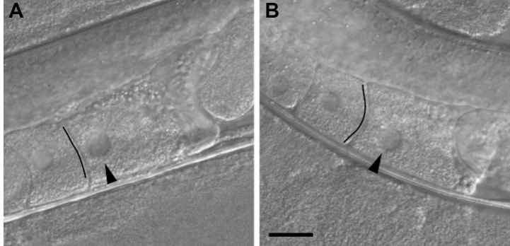
Figure 2. Inference of the timing of ovulation on the basis of the oocyte shape.
A. An oocyte that appears ready to be ovulated within a few minutes. The oocyte shape is rectangular, the boundary facing the next oocyte looks like a straight line (indicated by the black line), and the nucleus is close to the cortical region distal to the spermatheca. B. An oocyte not ready for imaging. The boundary has an arc-like appearance (indicated by the black line), and the nucleus is not close to the distal cortex. Differential interference contrast images of non-injected wild-type worms are shown. Images are flipped to be in the same direction as the confocal microscopy images in Figure 3. Arrowheads indicate the nucleus. The black lines were drawn to emphasize the boundaries. Scale bar = 20 µm.

