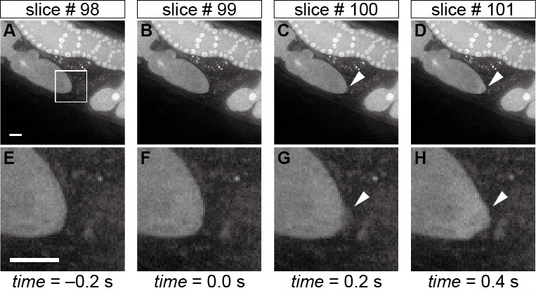
Figure 3. Morphological changes in the oocyte tip upon sperm entry.

Time-lapse images of the oocyte shape during fertilization in the whole field-of-view (A-D) and in the corresponding magnified region (E-H) indicated by the white box in panel A. The arrowheads in panels C, D, G, and H indicate the sudden protrusion of the oocyte tip upon sperm entry, and hence slice #99 is defined as the time 0 slice. Scale bars in panels A and E = 10 µm.
