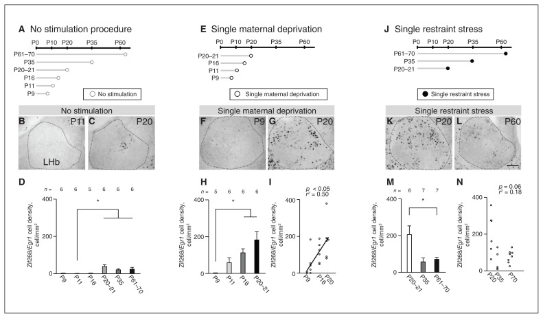
Figure 2.
Postnatal changes in neuronal reactivity in response to single maternal deprivation or single restraint stress during maturation of the LHb. (A) Schematic diagram of experiments without stress stimulation. (B and C) Zif268/Egr1 immunopositive cells at P11 and P20 with no stimulation. (D) Cell densities at each postnatal day. (E) Schematic diagram of experiments with single maternal deprivation. (F and G) Zif268/Egr1 immunopositive cells at P9 and P20 after single maternal deprivation. (H) Cell densities at each postnatal day. (I) Correlation between postnatal day and cell density after single maternal deprivation using a simple regression test. (J) Schematic diagram of experiments with single restraint stress. (K and L) Zif268/Egr1 immunopositive cells at P20 and P60 after single restraint stress. (M) Cell densities at each postnatal day. (N) Using a simple regression test, we observed no correlation between postnatal day and Zif268/Egr1 immunopositive cell density after single restraint stress. Scale bar = 100 μm. Error bars represent standard error of the mean. The square correlation coefficient (r) and p values of the slope are shown. *p < 0.05, Steel–Dwass multiple comparison test. LHb = lateral habenula; P = postnatal day.

