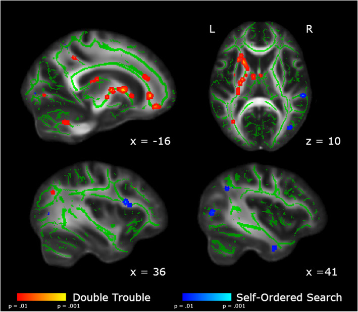
FIGURE 5.

Areas showing group differences in the change in FA from Baseline to Scan 5. In red, we show the areas in which the changes from Scan 1 to Scan 5 are significantly larger in the Double Trouble training group than in the Self‐Ordered Search training group. In blue, we show the areas in which the changes from Baseline to Scan 5 are significantly larger in the self‐ordered search training group than in the double trouble training group. Changes uniquely associated with Double Trouble were largely within the left inferior occipitofrontal and longitudinal fasciculi, while changes associated with Self‐Ordered Search were largely within the right superior longitudinal fasciculus. Clusters have been thickened for visualization using tbss_fill, and results are overlaid on the FMRIB58_FA template and the mean skeletonized FA data of the current sample
