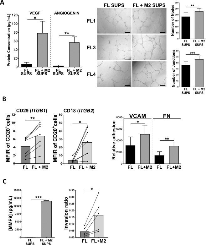
Fig. 3. M2 macrophages increases FL cells angiogenesis, adhesion, invasion, and related cytokine secretion.
A VEGF-A and angiogenin were quantified in supernatants (SUPS) from FL monocultures and FL-M2 co-cultures (48 h; left panels). Those supernatants were also used in a HUVEC tube formation assay (right panels). The number of nodes and junctions per field were obtained using ImageJ software with Angiogenesis Analyzer plugin. The data is representative of 3 FL patients. B Surface expression of CD29 (Integrin beta-1, ITGB1) and CD18 (Integrin beta-2, ITGB2) was analyzed by flow cytometry in FL primary samples (n = 7) after 48 h of culture with or without M2 (left panels). FL cells from FL monocultures or FL-M2 co-cultures (48 h) were subjected to an adhesion assay to VCAM or Fibronectin (FN) (n = 5) (right panel). C MMP9 vas quantified in supernatants (SUPS) from FL monocultures or FL-M2 co-cultures (n = 5) and the corresponding FL cells were recovered and subjected to an invasion assay for 24 h.

