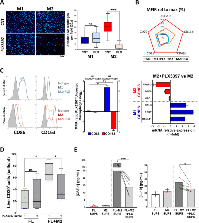
Fig. 6. CSF-1R blockade with PLX3397 inhibits M2 macrophage adhesion and reprograms M2 macrophages to M1, disrupting crosstalk between FL cells and M2 macrophages.
A M0 macrophages were detached and pre-treated for 1 h with 50 nM PLX3397, then seeded for 24 h in the presence of CSF-1 + IFNγ + LPS in the case of M1 macrophages or CSF-1 + IL4 for M2 macrophages. Attached macrophages were stained with Hoechst 33342 and quantified by Image J. B CSF-1R and integrin expression profile of M1 and M2 macrophages after PLX3397 (50 nM, 48 h) treatment. C M1 and M2 selected markers were analyzed by flow cytometry (CD86 and CD163) and RT-qPCR (PPARG, TGFB, MRC1, CCL5, CCR7, and CXCL11) in M2 macrophages treated or not with PLX3397 (50 nM, 48 h). D Primary tumor cells from FL patients (n = 4) were treated with PLX3397 (50 nM, 48 h) in co-culture or not with M2 macrophages (CSF-1 + IL4). Total number of live (LIVE/DEAD Aqua−) CD20 + cells is shown. E Concentration of CSF-1 and IL-10 determined by ELISA in supernatants from 6F. Statistical significance was assessed using paired t test or Mann–Whitney test.

