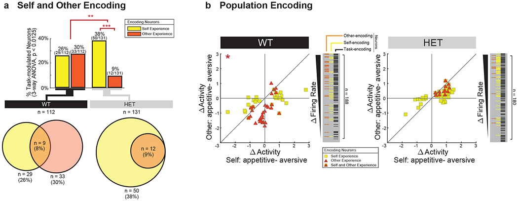
Fig. 2. Diminished Shank3 expression leads to diminished neuronal encoding of other-experience and diminished distinction between other-and-self.

a. Proportion of task-modulated neurons that encoded for self- and other-experience. Differences in encoding characteristics within and between the WT and HET mice were made by Chi-square test analysis (** χ2(1) = 15.38, p=8.78x10⁻⁵, *** χ2(1) = 30.51, p = 3.32x10⁻⁸). Venn diagrams below display the degree to which neurons that encoded self- vs. other-experience overlapped. Supplementary Table 2 provides additional results based on decoding. b. Scatter plot indicating the differences in neural activity (z-score) on trials in which appetitive (positive) vs. aversive (negative) experiences were given and illustrating the magnitude of response across conditions for individual cells. Here, the y-axis reflects absolute differences in activity based on whether the other animal had an appetitive vs. aversive experience (i.e., other-experience) and the x-axis reflects absolute differences in activity based on whether the recorded animals themselves had an appetitive vs. aversive experience (i.e., self-experience). Therefore, a neuron that responded preferentially to the other’s experience would be found midline along the vertical axis of the graph whereas a neuron that responded preferentially to one’s own experience would be found midline along the horizontal axis of the graph. Plotted cells are those which displayed significant modulation (three-way ANOVA, p<0.0125) to self (yellow) and other (orange) experience. The overlap between encoding properties per cell can be seen within the scatter plots and the insets to their right. Differences in the distribution of neuronal responses to self- vs. other-experience were evaluated for by a 2D 2-sample KS test (* p < 0.001). n = 112 and n = 131 task-modulated neurons recorded from n = 12 WT and n = 12 HET mice, respectively.
