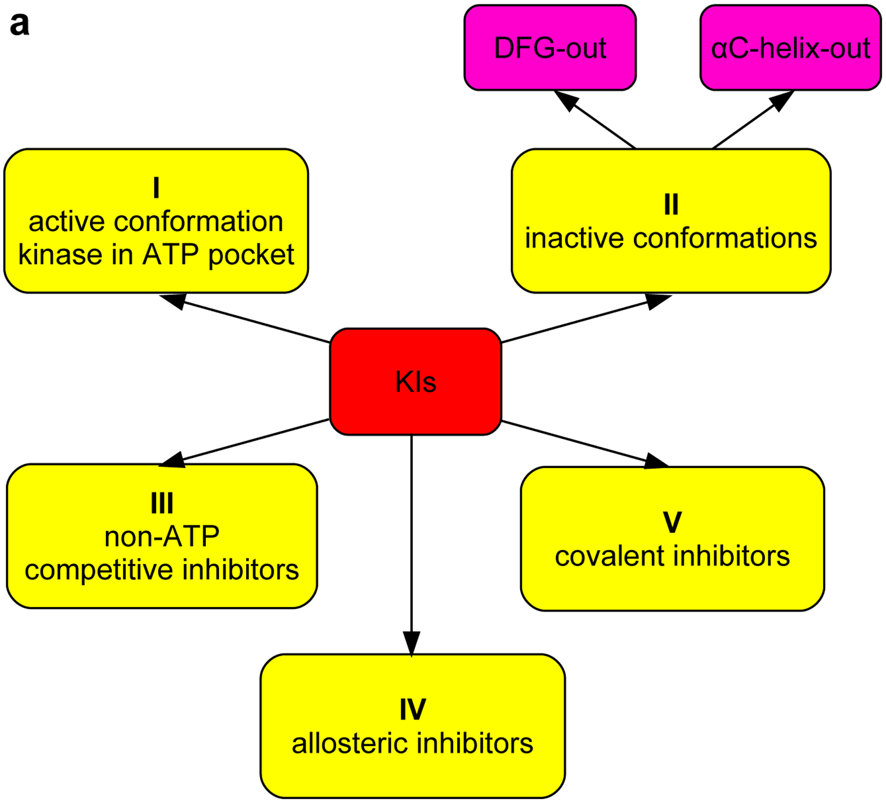
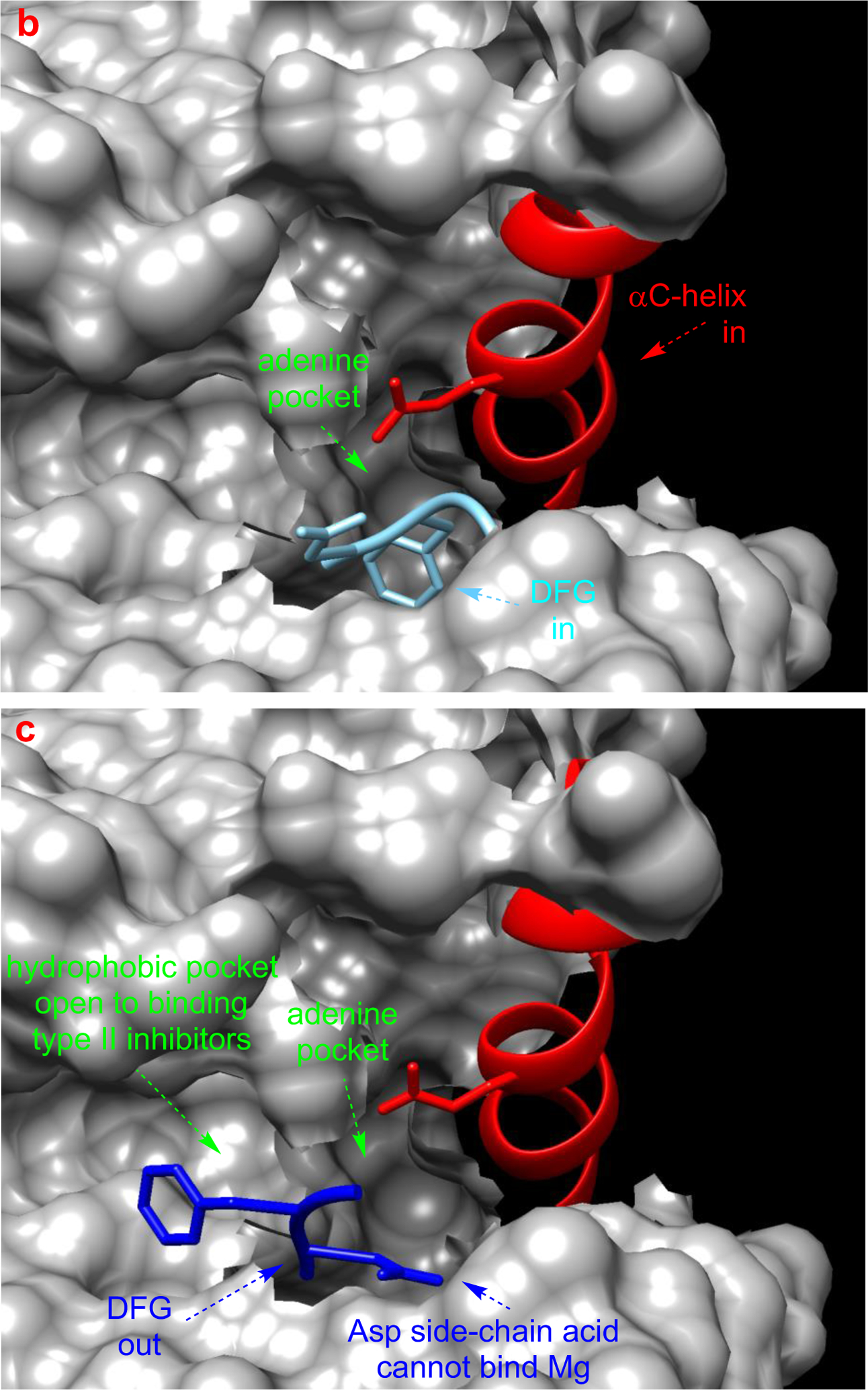
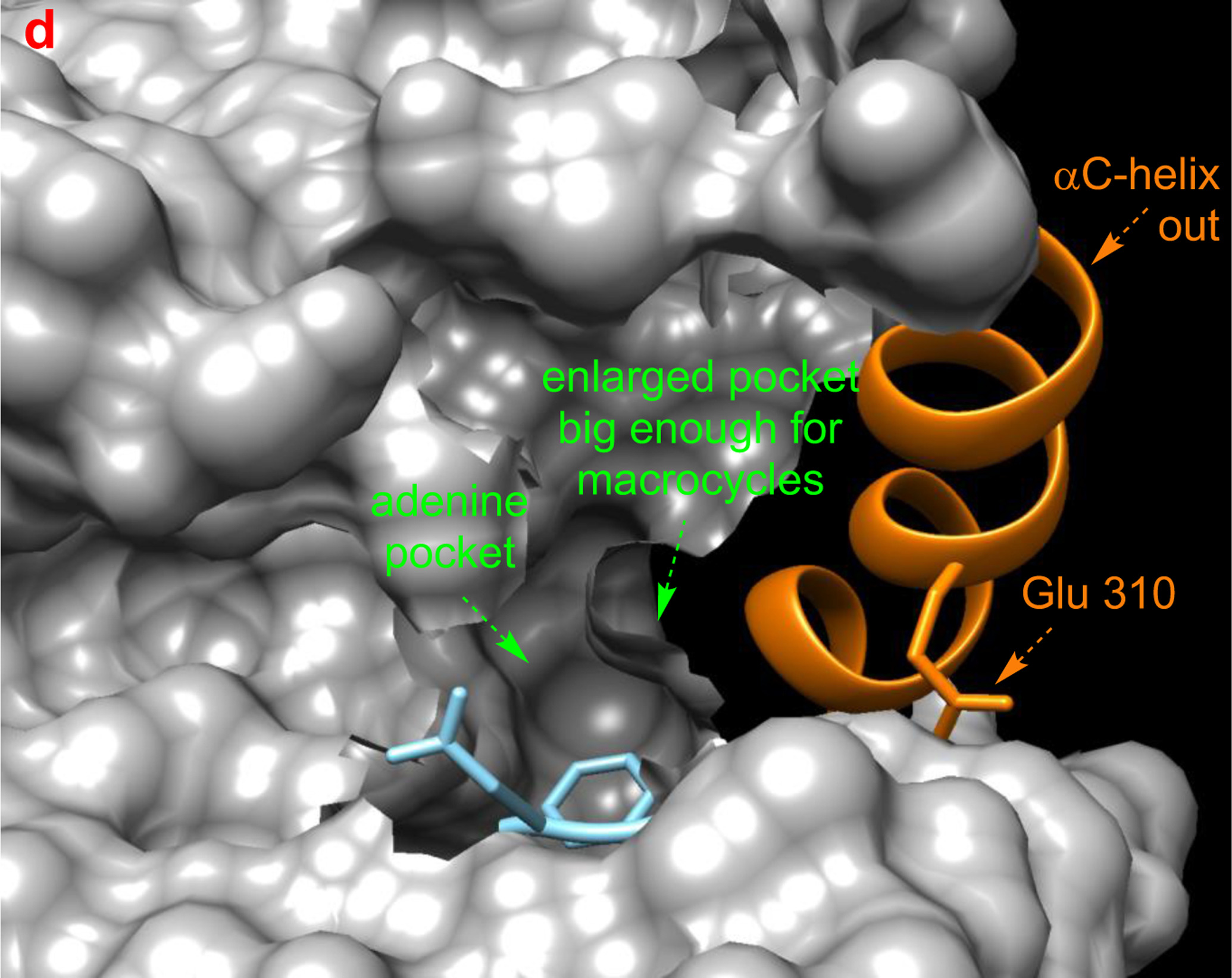
Fig 2.


a Different type of KIs. Structure of Src with kinase inhibitors in active (b, PDB: 3G5D), DFG-out (c, PDB: 4YBJ) and C-helix-out conformation (d, PDB: 4YBK). Type II inhibitors lead to DFG-out (dark blue) in contrast to DFG-in (light blue) conformation. c-Helix-out inhibitors lead to αC-helix-out (orange) in contrast to αC-helix in (red) conformation. Small molecule ligands are excluded for simplicity.
