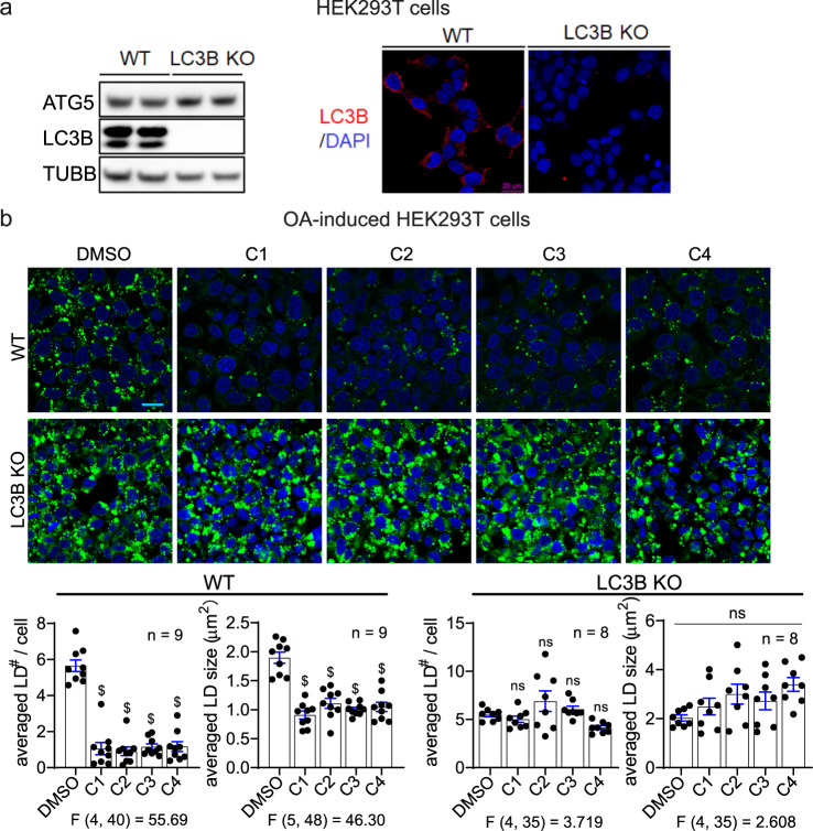
Fig. 5. LC3B knockout abolished the effects of LD·ATTECs.
a Representative LC3B western blot and immunofluorescence for the WT versus LC3B-knockout HEK293T cells. Note that different LC3B antibodies were used for western blot and immunofluorescence as indicated in “Material and Methods”. b Representative images and quantifications of the BODIPY493/503 staining of the OA-induced LDs in the WT versus LC3B-knockout HEK293T cells treated with the indicated compounds (5 μM for LD·ATTECs). Scale bar, 20 μm. The LD number per cell and averaged LD size in each field were quantified by ImageJ (particle analysis) in a blinded manner. n indicated the number of independently plated wells from two batches of experiments. ns, P > 0.05; $P < 0.0001 by one-way ANOVA (the F and degree of freedom values have been indicated) and Dunnett’s post hoc tests (compared to the DMSO group).

