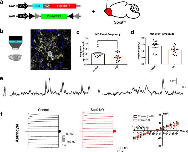
Fig. 2. Sox9 deletion disrupted cell intrinsic properties in astrocytes.
a Schematic of virally-induced Sox9 KO approach and targeted expression of GCaMP in astrocytes with Sox9 deleted. b Schematic of two-photon imaging of GCaMP6m from an ex vivo slice from Sox9fl/fl animals injected with a GFAP-driven Cre virus. Images were captured at ~1 Hz over 5 min. Approximate astrocyte territory (outlined in blue) defined the range for a single astrocyte and contained multiple astrocyte microdomains (outlined in yellow). Scale bar, 10 µm. c, d Quantification of calcium events in microdomains (MDs) of astrocyte processes. Astrocytes with Sox9 KO have a lower frequency of calcium events in microdomains but no change in amplitude. *p = 0.0435 Unpaired two-sided t-test. n = 12 cells from N = 3 control mice and n = 15 cells from N = 4 Sox9 KO mice. Data are presented as mean values ± SEM. e Example traces of GCaMP6m signal in microdomains of astrocytes in control and Sox9 KO astrocytes. f Whole-cell patch clamp electrophysiology of astrocytes show no changes in the I-V curve of astrocytes with stepped current injections in Sox9 KO vs controls. n = 10 cells (control) and n = 10 cells (Sox9 KO) from N = 3 mice, respectively. Data are presented as box plots displaying interquartile range and median with Tukey whiskers. Controls are C57BL/6 J injected with AAV-GFAP-iCre-P2a-TurboRFP + AAV-GFAP-flex-GCaMP6m.

