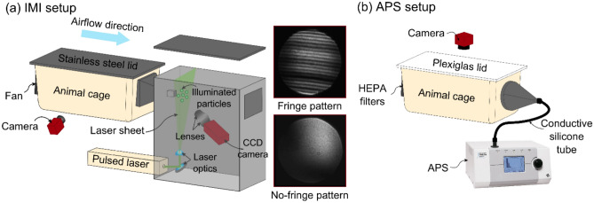
Figure 1.
Schematic of interferometric Mie imaging (IMI) and aerodynamic particle sizer (APS) setup. (a) IMI setup for counting and qualitatively characterizing particles emitted from a guinea pig cage. Particles emitted from the cage are carried toward the laser sheet by a horizontal airflow. A CCD camera captures out-of-focus images of the illuminated particles; images at right show representative water droplets with diameter of > 2 µm (fringe pattern), and < 2 µm (no-fringe pattern). A second camera captures the time-lapse images of guinea pig in the cage (side-view). (b) APS setup for measuring the size distribution of particles with diameter of 0.3–20 µm emitted from a guinea pig cage. An APS pulls the air at 5 L/min through the HEPA filters and a camera captures top-view images of the guinea pig in the cage at 1 image per second.

