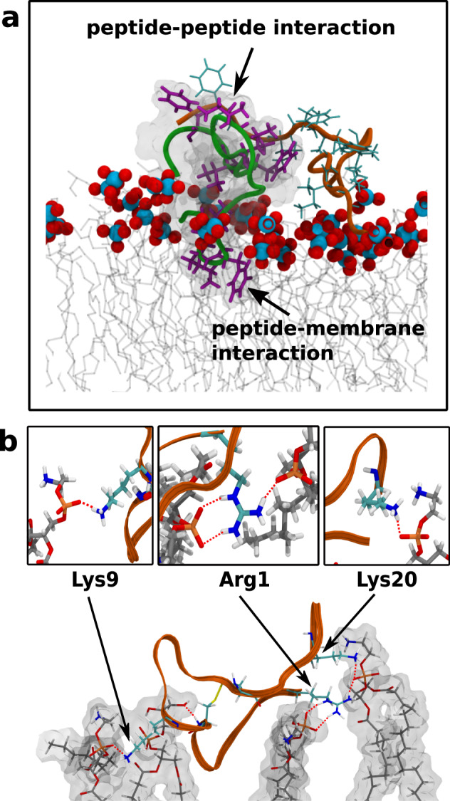
Fig. 9. Observed peptide-membrane interactions.

a Hydrophobic residues involved in peptide-peptide and peptide-membrane interactions. Two peptides (in green and orange ribbon) and their respective hydrophobic residues are depicted (in magenta and cyan sticks). Blue-red balls denote lipid phosphate groups, while the acyl chains are depicted in gray lines. b Polar interactions between HSEP3 peptide (ribbon in orange) and the membrane lipids (sticks) as observed in the final snapshot of simulation. The h-bond interactions (red line, dotted) are chiefly mediated by the cationic residues—lysine and arginine.
