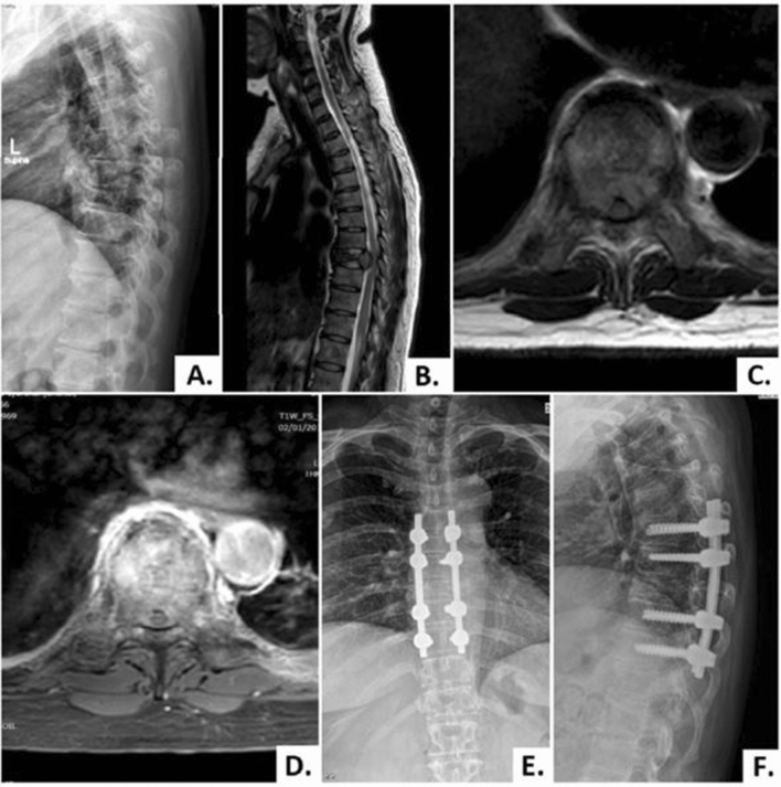
Figure 2.
A 49-year-old female patient with breast cancer (case No. 15). (A, B) Lateral plain radiograph and sagittal T2-weighted MRI showed the collapse of the T8 vertebra and metastases tumor infiltration at the vertebral body with extension posteriorly into the spinal canal. (C, D) Axial T2-weighted and T1-weighted contrast-enhanced MRI showing enhancement of the vertebral body and both sides of pedicles with the metastatic tumor extension already encased and compressing the spinal cord. (E, F) Postoperative anteroposterior and lateral plain radiograph obtained at 3-month follow-up visit showing proper implant position without loss of fixation.

