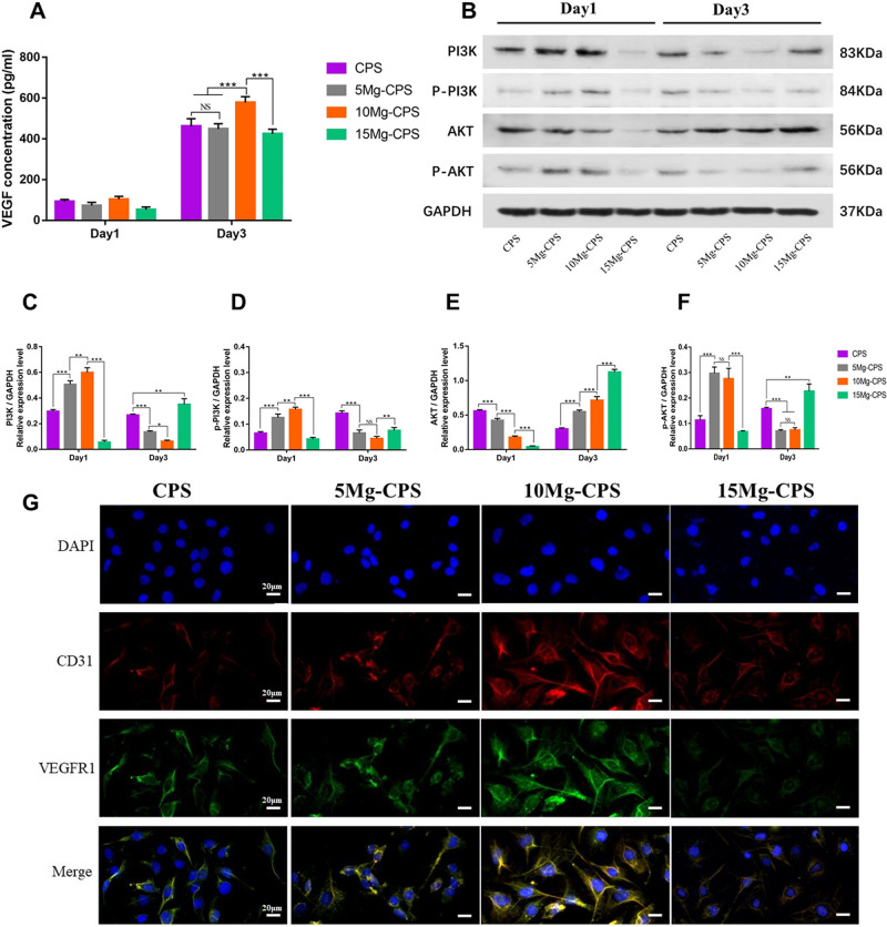
Figure 6.
Expression and secretion of proangiogenic cytokines stimulated by extracts. (A) Exhibited the concentration of VEGF secreted by HUVECs in extracts on day 1 and day 3. (B) Showed the levels of PI3K, phosphorylated PI3K (p-PI3K, activated form of PI3K), AKT, phosphorylated AKT (p-AKT, activated form of PI3K) and GAPDH of HUVECs on day 1 and day 3. (C–F) Illustrated the average ratios of PI3K/GAPDH (C), p-PI3K/GAPDH (D), AKT/GAPDH (E) and p-AKT/GAPDH (F) based on the intensity analysis of gray bands. (*P < 0.05, ** P < 0.01, ***P < 0.001, NS = not statistical). (G) Presented the immunofluorescence of 4’,6-diamidino-2-phenylindole (DAPI) staining, anti-CD31 antibody, anti-VFGFR1 antibody and the merge of them (scale bar =20 μm)

