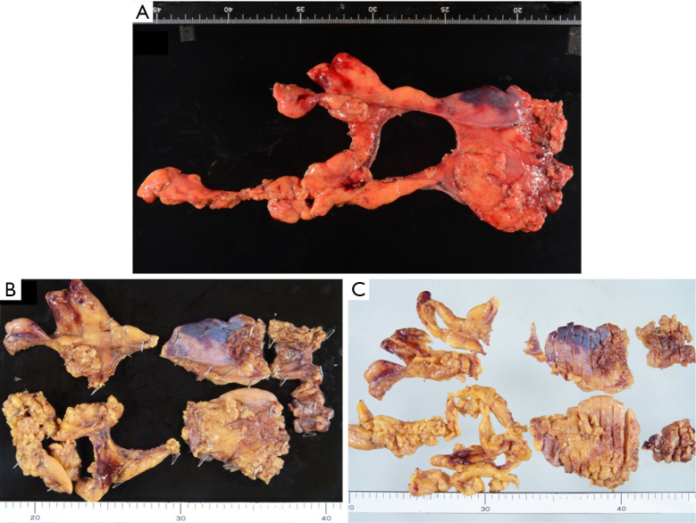
Figure 1.
Thymic tissue (case 3) resected by extended thymectomy (A). The thymic tissue was divided into 6 parts (upper right lobe, upper left lobe, lower right lobe, lower left lobe, right pericardial fat tissue and left pericardial fa tissue) and embedded in paraffin blocks after fixed in 10% formalin (B). Tissue blocks were sliced (C) and stained using hematoxylin and eosin.

