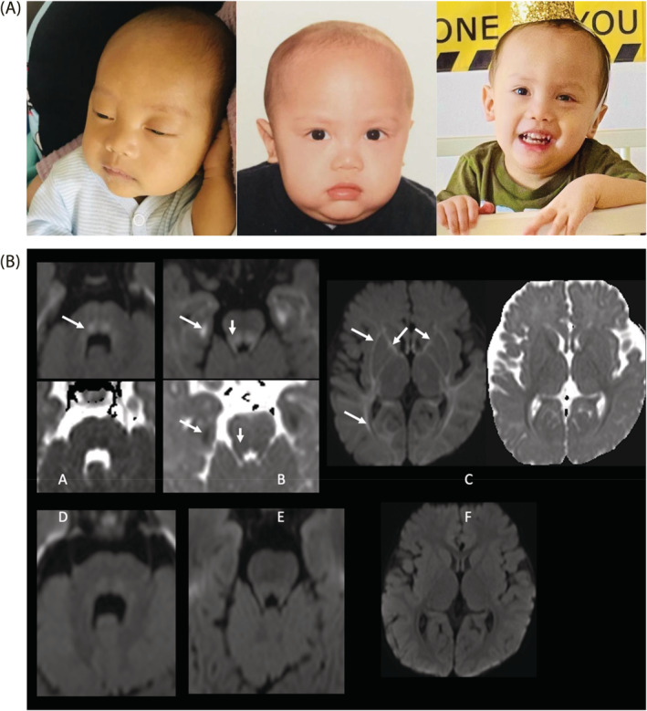
FIGURE 2.

Dysmorphic features and brain MRI findings. A) Picture on the far left shows our patient at 3 months of age with jaundice, frontal bossing, macrocephaly and coarse face. Picture in the middle shows our patient at 11 months of age after initiation of low‐methionine diet. We can see the same dysmorphic features in addition to body‐built improvement following weight gain. Picture on the right is his second birthday, hypotonia can be appreciated in the hands. B), Brain MRI finding showing diffusion imaging (b = 1000) and ADC map at 6 (A, B, C) and 12 (D, E, F) months of age. Panel A showed descending dorsal fibers of the tegmentum with high signal (white arrow) that resolved at 12 months of age (Panel D). Panel B shows restricted diffusion in the mesial temporal subcortical white matter as well as in the tegmentum and superior cerebellar peduncles (white arrows) that resolved at 12 months of age (Panel E). Panel C shows mild restricted diffusion in the supratentorial white matter involving the internal and external capsules, optic radiations and subcortical white matter (white arrows) that resolved at 12 months of age (Panel F)
