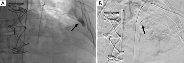
Figure 7.
ICA angiogram demonstrating (A) tortuous path of the ICA along with the site of active bleeding (arrow) in patient who developed hemorrhagic shock 10 hours post ultrasound guided left thoracentesis. (B) The ICA was successfully embolized (arrow) and no flow was demonstrated across the injury site. ICA, intercostal artery.

